
Dog diseases · Bones, joints and muscles
Bone cancer in dogs
Bone cancer can cause persistent lameness or a painful, hard swelling in a limb. Osteosarcoma is the most common malignant tumour originating in canine bone.
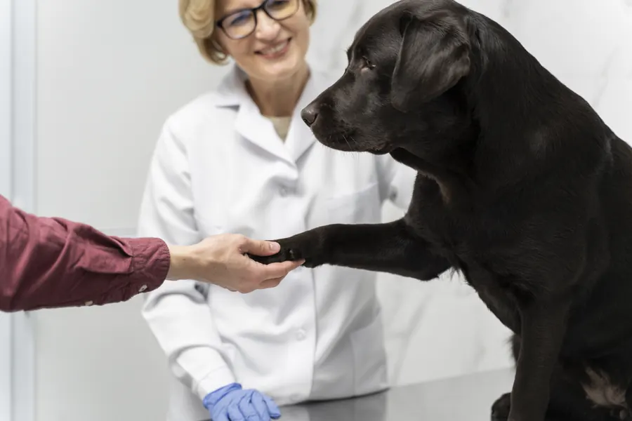
When to see a vet urgently
Arrange an appointment soon for persistent or worsening lameness or hard, painful limb swelling. Call an emergency clinic immediately if your dog suddenly stops using a limb or pain becomes severe.
- Limb position changes suddenly with severe pain.
- Your dog cannot stand without support.
- Breathing becomes laboured.
- General condition deteriorates rapidly.
Overview
Osteosarcoma and other bone tumours
Osteosarcoma involves uncontrolled growth of cells in bone-forming tissue. It accounts for over 80% of canine bone tumours. Limb bones contain 69–86% of osteosarcomas. Other sites include the lower jaw, ribs and vertebrae.
Chondrosarcoma is the second most common malignant tumour originating in canine bone. Fibrosarcoma can also arise in bone tissue. Cancers elsewhere can metastasise to bone. Benign tumours such as osteoma are rare in dogs.
Limb tumours are often found at the lower radius near the wrist. The upper humerus near the shoulder is another common site. Hind limb sites include the lower femur or upper tibia near the knee.
A tumour can also grow directly into bone from adjacent soft tissue.
Causes
Tumour development and growth
Most osteosarcomas start within the medullary cavity. Origin in the periosteum, the bone's outer covering, is less common. Limb tumours often lie between a long bone's end and shaft. Tumours can destroy normal architecture while new bone forms around them. Some large breeds have inherited susceptibility.
Tumour cells can contain alterations in genes normally limiting cell growth. Individual cases have occurred at old, healed fracture sites. Tumours have also been reported in chronically inflamed bone.
Osteosarcoma occasionally occurs near orthopaedic implants, but this is very rare.
Risk factors
Large dogs, including Great Danes and Rottweilers, are affected more often. In UK primary care in 2016, 0.037% of dogs had osteosarcoma. Annual prevalence in Scottish Deerhounds was 3.28% in the same study. Leonbergers had 1.48%. Great Danes had 0.87%. Rottweilers had 0.84%. Median diagnosis age was 9.64 years.
One breed health report from northern Europe describes typical onset in Irish Wolfhounds at four to six years. Tumours occasionally develop in young dogs nearing the end of growth. Small dogs can also develop lower jaw, rib or pelvic osteosarcoma.
Greater height at the withers is associated with increased risk alongside large size.
Typical symptoms
Limb osteosarcoma can start as lameness worsening with growth of the tumour. Signs depend on the affected bone.
- Your dog may protect the painful leg while walking.
- A hard, localised swelling can develop over the bone.
- Severe pain may stop your dog eating.
- Pain can change normal behaviour.
- Lameness can worsen suddenly if weakened bone fractures.
Jaw tumours can make eating difficult. Jaw osteosarcoma can cause swelling. Opening the mouth may become difficult. Drooling can increase. Vertebral tumours can cause an unsteady gait.
Muscles in the painful limb can shrink with reduced use. Dogs may tire sooner on walks. Pain can make a comfortable resting position hard to find.
Complications and consequences
Fractures, spread and prognosis
Tumour-damaged bone can fracture without major trauma. Osteosarcoma often metastasises early. The lungs are a common metastatic site. Detectable metastases were found at diagnosis in around 12% of dogs.
Severe pain or fracture limits most untreated dogs' survival to around one to two months. Local limb tumour treatment without chemotherapy has produced median survival of around four to 5.8 months. Amputation alone has an estimated median of four to 6.5 months. Amputation with chemotherapy gives a median of around nine to 12 months. Survival with surgery and chemotherapy has also been estimated at 12–14 months. Around 40% were alive at one year after local treatment plus chemotherapy. Around 20% receiving the same combined approach were alive at two years. Chondrosarcoma can have a better prognosis, with an estimated median of one to three years after wide excision.
Osteosarcoma can spread to other bones as well as lungs. Incomplete removal of skull, spinal or rib tumours can allow local recurrence. Raised blood alkaline phosphatase, or ALP, is associated at group level with shorter survival in limb osteosarcoma.
One follow-up after precisely targeted radiotherapy found fractures of weakened bone in 41% of cases. Most dogs' lameness improved within three weeks in that follow-up.
Diagnosis at the vet
Imaging and sampling the lesion
The vet identifies which bone is painful or swollen. X-rays are usually taken of the suspected area. Local bone destruction can be visible. New bone around the periosteum can appear on the same images. Bone infection can resemble a tumour.
Fine-needle samples can reveal tumour cells. Biopsy includes extracellular material alongside cells, helping identify type. Imaging guides sample site selection because sampling can weaken bone enough to fracture.
A removed tumour can be examined in the laboratory for classification. After limb-sparing surgery, margins are also assessed for tumour extension.
Assessing spread
Three chest X-rays from different directions can look for visible metastases. Chest X-ray-visible metastases occur at diagnosis in around 5% of dogs with limb osteosarcoma. Chest CT detects spread more sensitively than X-rays. Apparently normal lungs can still contain microscopic metastases. A nearby lymph node can be sampled when assessing spread. Blood ALP can also rise in other bone lesions.
CT can map local skull, spinal or rib tumour extent before treatment decisions. A blood count assesses general health before anaesthesia or chemotherapy planning. Abdominal ultrasound can seek a primary tumour if bone metastasis from another cancer is suspected.
Treatment
Tumour removal and chemotherapy
Amputation removes the bone tumour and source of local pain. Beforehand, the vet assesses whether remaining limbs can support body weight. Rib tumours may sometimes require removal of only the affected bone section. Limb-sparing surgery can suit selected dogs with appropriately located tumours. Its aim is to preserve affected limb function. Surgical-site infection can develop afterwards. Most dogs can move within 24 hours of amputation. Suitable supports can help immediately after surgery. Adapting to normal three-legged movement often takes around four weeks. Chest imaging every two to three months may be recommended to monitor metastases. Chemotherapy aims to delay metastatic development after local treatment.
Pain relief and treatment side effects
When surgery is impossible, care may aim to relieve pain and preserve function. NSAIDs can form part of veterinary tumour pain management. Progressing disease may need several medicines together. Radiotherapy can relieve tumour-related bone pain. Bisphosphonates, which slow bone breakdown, can form part of the pain plan.
Chemotherapy-associated nausea or vomiting can start within 24 hours. Delayed nausea or vomiting can begin one to five days afterwards. Blood tests can monitor neutrophils when chemotherapy may reduce these infection-protecting white cells.
Vomiting or diarrhoea can sometimes cause enough dehydration to require hospitalisation.
Prevention
The precise cause is unknown, so there is no certain preventive measure. Breed monitoring gathers information on cancers appearing only in adulthood. Early examination of persistent lameness or swelling in susceptible breeds helps identify the pain's cause. Some breed health programmes monitor osteosarcoma as a major Scottish Deerhound health concern.
Some Scottish Deerhound breed clubs also collect owner-reported illness information. Irish Wolfhounds may develop bone cancer only after they have already been bred.
Important for dog owners
Limit exercise to brief lead walks until the limb is examined. Avoid jumping and stairs while it is painful and awaiting assessment. Monitor whether your dog can rest peacefully and rise as usual. Contact the vet promptly for wound discharge or marked swelling after surgery.
After chemotherapy, promptly report fever or deterioration. Describe when vomiting or diarrhoea began relative to the last treatment.
Follow discharge precautions: exposure to bodily waste should be avoided for 72 hours after chemotherapy. If cleaning is needed, wear gloves and a mask as the treating clinic instructs.
Frequently asked questions
Can bone cancer occur without a visible lump?
A limb tumour may initially cause only intermittent lameness or reluctance to move. Hard swelling can become apparent later. Persistent lameness may prompt imaging of the painful site even without a clear lump.
Can X-rays alone diagnose osteosarcoma?
Typical changes can raise suspicion. Bacterial or fungal bone infection can look similar. Biopsy helps distinguish tumour from other lesions and identify its type.
Why image the lungs without a cough?
Limb osteosarcoma can spread to lungs early. Imaging seeks visible metastases before selecting treatment. Microscopic spread can remain invisible despite normal X-rays.
Can dogs manage on three legs?
Many adapt and gradually resume normal activities. Disease in remaining limbs can make adaptation harder. Supports are available for dogs needing help on initial post-operative walks.
What if the tumour is not operated on?
The vet can plan pain medication even without removal. Radiotherapy can sometimes relieve bone pain. Movement, eating and resting at home help assess whether relief is adequate.
Sources
- Osteosarcoma of the appendicular skeleton in dogs: consensus and guidelines, Frontiers in Veterinary Science, accessed 2026-11-07.
- Dog breeds and conformations predisposed to osteosarcoma in the UK, Canine Medicine and Genetics, accessed 2026-11-07.
- Bone Tumors in Dogs and Cats, Merck Veterinary Manual, accessed 2026-11-07.
- Canine Bone Tumors, North Carolina State University College of Veterinary Medicine, accessed 2026-11-07.
- Osteosarcoma in dogs, Cornell University College of Veterinary Medicine, accessed 2026-11-07.
- Emergency and Critical Care: emergent conditions, Cornell University College of Veterinary Medicine, accessed 2026-11-06.
- Mitä on päivystyshoito, Helsingin yliopisto, accessed 2026-11-07.
- Koiran ontuminen, FirstVet, accessed 2026-11-07.
- Koiranjalostuksen ongelmat ja valvontakriteerit, Luonnonvarakeskus, accessed 2026-11-07.
- Tunnistatko koirasi kivun?, Suomen Kennelliitto, accessed 2026-11-07.
- Kotihoito-ohjeet toimenpiteen jälkeen, Kuopion Eläinlääkärikeskus, accessed 2026-11-07.
- Irlanninsusikoiran jalostuksen tavoiteohjelma 2025–2029, Suomen Irlanninsusikoirat, accessed 2026-11-07.
- Skotlanninhirvikoira, Suomen Kennelliitto, accessed 2026-11-07.
- 2026 AAHA Oncology Guidelines for Dogs and Cats, American Animal Hospital Association, accessed 2026-11-07.
- Bone Tumors in Cats and Dogs, American College of Veterinary Surgeons, accessed 2026-11-07.
- Osteosarcoma in Dogs, VCA Animal Hospitals, accessed 2026-11-07.
This information does not replace a visit to a vet. In an emergency, contact your nearest emergency vet: find a vet (RCVS).
More on this topic: Bones, joints and muscles
- Atlantoaxial instability in dogs: neck pain and treatment
- Crooked front legs in puppies: angular limb deformity
- Cruciate ligament injury in dogs – a torn cranial cruciate ligament
- Hip dysplasia in dogs: symptoms, tests and treatment
- Immune-mediated polyarthritis in dogs: symptoms and care
- Tendonitis in dogs – symptoms, investigations and treatment