
Dog diseases · Eyes, ears and mouth
Epulis in dogs – assessment and treatment of gum lumps
Epulis is a general term for a growth rising from the gum, rather than a precise diagnosis. One such lesion is a peripheral odontogenic fibroma, or POF. However, a similar lump may be an ameloblastoma that destroys bone locally. A tissue sample helps identify the lesion and select treatment.
When to see a vet urgently
A mouth lump can interfere with eating and cause pain.
- There is heavy or continuous bleeding from the mouth.
- Your dog cannot chew or keeps dropping food.
- Closing the mouth is difficult and your dog seems painful.
- Facial swelling increases and your dog resists having the head touched.
These signs need veterinary assessment without delay. The vet guide can help you find assistance.
Overview
The name epulis does not establish whether a lesion is simply excess tissue, a benign tumour or a locally aggressive tumour. The older terms fibromatous and ossifying epulis usually refer to POF and its bone-forming variant.
A peripheral odontogenic fibroma is usually a benign, firm tissue growth near the teeth. POF is among the most common benign oral tumours in dogs.
It often occurs towards the front of the upper jaw near the incisors or canine teeth. Acanthomatous epulis is now known as canine acanthomatous ameloblastoma, or CAA.
CAA can invade neighbouring jawbone despite being classified as a benign tissue type. Local fibrous hyperplasia associated with inflammation or irritation is a separate diagnosis.
Reliable distinction between POF and local fibrous hyperplasia requires histological examination.
Causes
The tissue of origin varies
The precise tissue from which POF arises has not been definitively established. Suggested origins include gum cells, surface epithelium, tooth socket bone and the periodontal ligament.
Benign reactive gum hyperplasia can instead develop after inflammation or injury. Boxers also develop inherited, widespread fibrous gingival hyperplasia.
No specific inherited genetic defect has been established as the cause of POF. An ameloblastoma is a tumour arising from epithelial tissue involved in tooth development.
Reactive gingival hyperplasia may be localised or widespread, with tissue covering the tooth's visible crown.
Bone formation and destruction
Some POF lesions produce bone within the tumour. Imaging of an ossifying POF shows bone formation, rather than the possible bone loss caused by CAA.
An ameloblastoma may begin within bone or grow in the gum's soft tissues. A tumour beginning within bone can particularly expand destructively through the jawbone.
Risk factors
Typical POF lesions mainly affect middle-aged and older dogs, often over six years old. A diagnostic laboratory's 2019 samples identified 1,001 confirmed POF diagnoses.
POF accounted for 2.8% of all canine biopsies at the laboratory. The average age of diagnosed dogs was 8.1 years.
The odds ratio for diagnosis in Boxers compared with crossbreeds was 3.78. For Border Terriers, the corresponding odds ratio was 3.21.
For Basset Hounds, it was 3.18. The laboratory mainly served first-opinion and referral practices in Britain and Ireland.
Multiple POF lesions were found simultaneously in 150 dogs. These figures describe diagnoses in submitted laboratory samples, rather than disease percentages for all dogs or other national populations.
Typical symptoms
POF may begin as a small gum lump and gradually enlarge.
- A firm, projecting lump is visible on the gum or between teeth.
- It may be smooth or cauliflower-like, and gum-coloured, pale or red.
- Your dog drools more or has blood-stained oral discharge.
- Eating looks difficult or food falls from the mouth.
- A tooth changes position or falls out.
- Weight loss may accompany an oral tumour.
- Your dog does not want the head or mouth area touched.
- Breath may smell worse than before.
- Swelling on one side of the face may alter the muzzle's appearance.
A small oral lesion may be found during examination before other symptoms are noticed at home. A lump is usually solitary, but several separate lesions can occur in the same dog.
Adjacent growths can merge and resemble widespread gum thickening. Of 85 measured lesions in the biopsy study, 78 were 5–50 millimetres in diameter.
A benign POF can look actively growing and displace teeth.
Complications and consequences
Distinguishing local growth from metastasis
Typical POF does not invade the jawbone or metastasise. However, growth can push teeth out of their normal positions.
A pocket-like area around a tooth may predispose it to periodontal disease. A large gum lesion can be injured during chewing or mouthing.
Local growth of CAA within the jawbone can cause severe bone pain. However, CAA does not usually metastasise elsewhere in the body.
Malignant transformation has not been described with recurrence of ordinary POF. Complete removal usually cures POF permanently.
Diagnosis at the vet
Whole-mouth examination and a tissue sample
The assessment also establishes the lump's growth rate and any tenderness, bleeding or difficulty eating. Other oral findings, such as tartar and possible periodontitis, are recorded as background information.
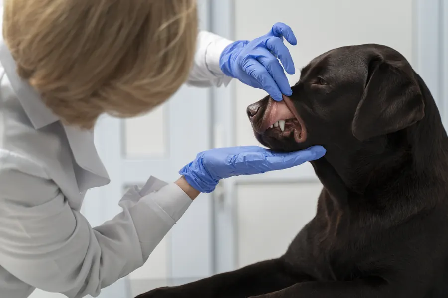
The vet examines the mouth and palpates the lesion and regional lymph nodes. A thorough oral examination may require sedation.
Appearance alone cannot confirm an oral tumour's type. A biopsy takes part of the lesion for microscopic examination.
This is called histopathology, examination of tissue structure. POF, reactive gingival hyperplasia and early CAA can look very similar to the naked eye.
A malignant tumour is another possible diagnosis for a mouth lump. An overly superficial sample may contain normal gum and miss a deeper growth.
Invasion of surrounding tissue is assessed more reliably when the biopsy includes enough deep tissue. If an X-ray shows a bone lesion, the vet may sample down to the bone to establish its nature.
A small, stalked lump can sometimes be removed completely for tissue examination at the same time. The pathologist benefits from knowing its location and whether the sample contains the whole lesion or only part.
Dental imaging and surgical planning
Dental X-rays assess whether underlying bone is affected. An ossifying POF may appear as a mineralised area within soft tissue.
CT helps establish tumour boundaries and the extent of bone changes. Imaging before surgery helps plan adequate removal at the first operation.
Depending on the tumour type, lymph nodes and the chest may also be assessed for spread.
Treatment
POF removal and preserving the tooth
The vet prescribes treatment and any medication according to the tissue diagnosis, imaging and the dog's condition. Usual POF treatment is complete removal of the gum growth.
In some cases, it can be removed down to the periosteum without extracting the tooth. POF does not usually require the same healthy tissue margin as an ameloblastoma.
However, removal may be combined with tooth extraction and curettage of the socket. If a large gum defect remains, the tooth may need extraction or reconstructive tissue protection around it.
A healthy tooth needs at least two millimetres of gum tissue around it. Ossifying POF is treated on the same principles as ordinary POF.
However, its hard, bony tissue can make removal technically harder. Incomplete removal can lead to local recurrence.
CAA treatment may require jawbone removal
Locally invasive CAA may require removal of part of the lower or upper jaw. A healthy tissue margin of about one centimetre, including bone, is usually recommended.
A five-millimetre margin may be considered in certain limited situations. With ameloblastoma, the aim is to remove all abnormal bone so that no local growth remains.
Local recurrence after extensive jaw resection has been reported at below 5%. Surgery aims to remove microscopic tumour tissue as well as visible growth, leaving none at the wound margins.
If complete removal is impossible or margins are narrow, further treatment may improve local control. Ameloblastoma is sensitive to radiation, so radiotherapy can be an alternative to surgery.
One radiotherapy course used lasts three weeks. Bone pain can be relieved with radiotherapy and systemic pain relief chosen by the vet.
If curative local treatment is not possible, care may focus on relieving pain and preserving quality of life.
Recovery and mouth function
Many dogs adapt well to partial jaw removal and are ready to eat within hours. Appearance may change after jaw surgery even when the dog can eat and drink.
Possible consequences include drooling, altered jaw alignment, an opening between mouth and nose, or wound breakdown. A protective collar is usually needed for the first 2–3 weeks after major oral surgery.
During that time, rubbing the face on carpets or furniture may disrupt healing. After healing, dogs can usually return to playing, chewing toys and eating dry food.
Prevention
Regular oral checks during annual examinations and dental care can find a lesion before symptoms develop. Treating the pocket-like gum growth caused by POF reduces problems from abnormal tissue remaining around the tooth.
Bone imaging helps plan adequate removal and reduces the risk of an incomplete first operation. Treating gum inflammation and repeated injury is particularly important for reactive fibrous growth, which may arise from irritation.
Follow-up checks look for local recurrence before it becomes difficult to remove.
Important for dog owners
Record when you noticed the lump and whether it seems to be growing. Mention bleeding, changes in chewing and food falling from the mouth.
Ask for the precise diagnosis from the tissue report and keep it. Distinguishing epulis, POF and ameloblastoma helps explain why different dogs are offered operations of different extents.
A mouth lump's benign-looking surface alone cannot establish its tissue type.
Before discharge, confirm what your dog may eat and chew and how to monitor the wound. Contact the practice again if your dog cannot eat, bleeding continues or the wound appears to open.
Frequently asked questions
Is an epulis always cancer?
Epulis is not a precise tumour type, but a term for a gum tissue growth. Ordinary POF is benign and does not metastasise. A similar-looking ameloblastoma can still destroy local bone, so a tissue sample is needed to establish the correct diagnosis and choose treatment.
Must a tooth always be removed with an epulis?
A small POF can sometimes be removed completely while preserving the tooth. If removal leaves too large a gum defect, tooth extraction or a reconstructive periodontal procedure may be needed. Treatment extent therefore also depends on the healthy tissue remaining around the tooth.
Can an epulis recur after removal?
POF can recur locally if some lesion remains. Complete removal usually cures ordinary POF permanently. Follow-up checks are nevertheless useful to detect possible new growth early, before treatment becomes harder.
Can a gum lump be identified from a photograph alone?
POF, gingival hyperplasia and early acanthomatous ameloblastoma can look alike. A definitive tissue diagnosis requires histopathology. Dental X-rays and, when needed, more detailed CT also show whether underlying jawbone has changed.
Can a dog eat after jaw surgery?
Many dogs adapt well to partial jaw removal and can resume eating. However, more extensive surgery may alter appearance and cause problems such as drooling. After healing, many dogs can play again and eat dry food.
Sources
- Oral Tumors in Small Animals, MSD Veterinary Manual, accessed 2026-11-03.
- Oral Tumors – Peripheral Odontogenic Fibromas, VCA Animal Hospitals, accessed 2026-11-03.
- Oral Tumors, American College of Veterinary Surgeons, accessed 2026-11-03.
- Pattitilanne suussa?, Solumo, accessed 2026-11-03.
- Clinicopathological features of peripheral odontogenic fibromas in dogs and risk factors for their laboratory diagnosis, Journal of Small Animal Practice; ARFCV public deposit, accessed 2026-11-03.
- It Is Not Called an Epulis Anymore, Today’s Veterinary Practice, accessed 2026-11-03.
This information does not replace a visit to a vet. In an emergency, contact your nearest emergency vet: find a vet (RCVS).