
Dog diseases · Heart and blood vessels
Heart block in dogs: slow heart rate and treatment
Atrioventricular block, or heart block, disrupts electrical impulse conduction from the atria to the ventricles. A mild disturbance can cause no symptoms. Severe block can produce a very slow rate and fainting. Complete block also carries a risk of sudden death. It generally requires a permanent pacemaker.
When to see a vet urgently
Fainting and circulatory collapse warrant urgent veterinary assessment.
- Your dog collapses or loses consciousness, even if the episode passes quickly.
- Your dog cannot remain standing or rapidly becomes profoundly weak.
- Breathing is difficult or clearly laboured.
- Gums become very pale or bluish.
- A dog with a pacemaker starts fainting or becoming weak again.
Overview
The normal heartbeat signal begins in the sinus node and spreads first through the atria. At the atrioventricular node, it briefly slows so the ventricles can fill before contraction. It then travels into the ventricular conduction pathways.
A block can slow conduction, stop some impulses or prevent transmission completely. In complete block, subsidiary ventricular pacemakers can generate a slower replacement rhythm. The heart can therefore keep beating despite lost coordination between atria and ventricles. However, a slow rate alone does not prove atrioventricular block.
Causes
Conduction tissue damage or another disease
Fibrosis of conduction tissue is a common cause of severe block in older dogs. A cardiac tumour or atrioventricular node injury can also disrupt conduction. Myocarditis can damage electrical conduction and cause heart block.
Block has also been described with other heart muscle or valve disease. Electrolyte disturbances can affect electrical activity. High potassium, for example, may accompany a conduction disturbance. Sometimes the precise cause remains unknown despite thorough investigation.
Medicines and nervous system effects
Beta-blockers, calcium channel blockers and digoxin can slow atrioventricular conduction. Sedation or anaesthesia can also reveal a disturbance. Excessive medication can cause blocks even in an otherwise healthy dog.
Strong vagal nerve activity can slow both sinus node function and atrioventricular conduction. This may be reversible when the underlying disease is treated. In contrast, a slow sinus rhythm in an athletic dog can be normal.
Risk factors
Heart block occurs particularly in older dogs accumulating age-related conduction degeneration. An average patient age of about 11 years has been described. Reported predisposed breeds include Cocker Spaniels, Labrador Retrievers and German Shepherd Dogs. Entire females are also overrepresented.
The disease is not confined to old age: a published case describes complete block in a 2-year-old dog. Other heart muscle or valve disease influences tolerance of a slow rhythm. Diseases causing metabolic or electrolyte disturbances, such as Addison's disease, can predispose to abnormal conduction.
Typical symptoms
Mild block can be an incidental finding without signs noticed at home. In severe block, signs relate to excessively slow ventricular rate and inadequate circulation.
- Your dog suddenly faints or collapses.
- Normal walks or play cause earlier tiring.
- Your dog seems weak or reluctant to move.
- Heartbeats or arterial pulses feel unusually infrequent.
- Longstanding disease may involve heart failure and breathing difficulty.
Slow-rate symptoms often appear around 40 beats per minute or below. Onset also depends on duration and general health. In complete block, rate generally fails to increase normally with exercise or excitement.
Complications and consequences
Inadequate circulation strains the body
With an excessively slow rate, cardiac output may fall short of the body's needs. Weakness, reduced exercise tolerance and loss of consciousness can result. Severe conduction block can lead to heart failure or sudden death.
The heart can compensate by increasing the volume pumped with each beat. Atria and ventricles may consequently enlarge despite initially normal contractility. Enlargement can also stretch the mitral valve opening and cause regurgitation.
Treatment outlook and surgical risks
Pacing can reduce fainting and improve activity and quality of life. Among 52 dogs receiving an epicardial pacemaker on the heart's outer surface, 96% survived to discharge. Complete atrioventricular block was the indication in 39 dogs (75%).
Intraoperative complications were recorded in 11% of this group. Postoperative complications occurred in 23%. Concurrent heart muscle or valve disease was associated with shorter survival. Lead displacement may require another procedure. It most commonly occurs early in recovery, within the first days or weeks. Other possible problems include infection, lead damage and fluid accumulating around the device.
Diagnosis at the vet
ECG distinguishes the degrees of block
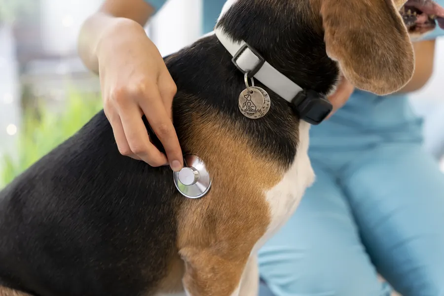
Your vet listens to the heart as part of a general examination. Assessment may also include blood pressure. An electrocardiogram, or ECG, shows whether atrial impulses reach the ventricles normally. The P wave represents atrial electrical activation and the QRS complex ventricular activation.
In first-degree block, all impulses conduct, but more slowly than usual. In second-degree block, some atrial impulses fail to reach the ventricles. In Mobitz type I, conduction time lengthens before a dropped beat. Mobitz type II lacks this progressive lengthening.
In high-grade second-degree block, several consecutive impulses fail to conduct. In third-degree block, no atrial impulse reaches the ventricles. Atria and ventricles then beat independently. Holter monitoring can detect intermittent block missed during a short ECG. It records electrical activity for at least 24 hours in ordinary daily surroundings.
Assessing the heart and possible underlying disease
Ultrasound assesses structure, contractility and valve changes. Blood tests may include a count, biochemistry and electrolytes. Depending on circumstances, thyroid function and troponin for heart muscle injury are also tested. Infection investigations may include tick-borne disease tests and tests for heartworm or Toxoplasma, for example.
An atropine response can help distinguish strong vagal activity from conduction tissue damage. Vagal slowing generally accelerates markedly with atropine. Complete block generally does not respond similarly. Before pacemaker implantation, anaesthetic risk and other diseases affecting long-term outcome are assessed. If heart failure is present, medication stabilises it before implantation. Atrioventricular valve degeneration does not necessarily prevent pacing, but severity is assessed first.
Treatment
Mild and reversible block
Mild, symptom-free first-degree block often needs no specific medication. Vagal Mobitz type I block can be physiological in a puppy. Mobitz type II needs monitoring because it can progress to more severe conduction disease.
Treatable electrolyte or metabolic abnormalities are corrected. Transient mild block during sedation may disappear when the drug's effect ends. The need for and suitability of medicines affecting rate are reviewed at the practice. Rate-increasing medication may help some second-degree blocks.
A pacemaker maintains rhythm in severe block
Complete block often involves permanent conduction tissue damage that medicine cannot repair. Pacing is then recommended even without symptoms because of sudden death risk. Symptomatic high-grade second-degree block may also need a pacemaker.
A pacemaker has a programmable pulse generator and connected cardiac lead. It senses the natural rhythm and delivers an impulse if ventricular rate falls below the programmed minimum. The impulse makes the ventricle contract and helps maintain adequate circulation.
The lead usually passes through a jugular vein into the right ventricle. Its tip can attach with a screw or passively engage structures on the ventricular inner surface. An epicardial pacemaker may be chosen for a small dog or after displacement of a previous lead. The generator is placed under skin around the neck or shoulder blades. Permanent implantation is performed under general anaesthesia. Severe bradycardia may require temporary pacing during the procedure.
Recovery, programming and follow-up
Exercise is generally restricted for about 30 days after implantation. This allows healing and reduces lead movement risk. Device function and battery status are checked after about 1 month.
Checks continue every 6 months, for example, or annually in a stable patient. Programming can adjust pacing rate and sensing sensitivity. Suitable settings can extend device life by several years. An activity sensor can increase rate during movement and improve exercise tolerance. With physiological pacing, ventricular rhythm follows atrial rhythm, allowing atrial contraction to aid filling as in normal sinus rhythm.
A pacemaker does not remove the possible medication needs of other heart disease. However, enlargement caused by a slow rhythm may decrease after pacing. In a follow-up of 3 dogs, pacing eliminated reduced exercise tolerance or fainting.
Prevention
Mobitz type II cannot always be prevented. Regular health checks can help find other conditions affecting cardiac function. Blood tests can identify treatable metabolic or electrolyte disturbances.
Watch for condition changes after starting a new medicine. Diagnosed second-degree block is monitored with repeat ECGs for progression. Mobitz type II may increase anaesthetic risk, so rhythm is monitored carefully during procedures. In pacemaker patients, avoid forceful pulling on the neck to help prevent lead displacement.
Important for dog owners
Record when fainting occurs and whether it happens at rest or during activity. Watch whether your dog manages their familiar walk as before. Mention previous heart block whenever your dog attends a new practice.
Keep the pacemaker wound clean and dry. Mention the device before MRI or CT. An ordinary microwave oven is not dangerous to a dog with a pacemaker.
New weakness or fainting warrants a device check before the scheduled appointment. Home activity monitoring helps identify changes but does not replace ECG.
Frequently asked questions
Does a slow rate always mean heart block?
A slow sinus rhythm can be normal in an athletic dog. Strong vagal activity can also slow rate. Sinus node dysfunction is another cause of bradycardia. ECG distinguishes atrioventricular block by showing the relationship between atrial and ventricular electrical activity.
Does a dog without symptoms need a pacemaker?
In complete block, pacing is recommended even without signs because of sudden death risk. Mild first-degree block, however, may have little significance. Mobitz type II is monitored for progression. Decisions use ECG-confirmed type and other cardiac findings.
Can block disappear after medication stops?
Reversible conduction disturbance associated with medication or anaesthesia may resolve when the effect ends. The same can happen after correction of a treatable electrolyte abnormality. Addison's disease is one possible underlying cause of such abnormalities. Complete block from permanent conduction damage generally needs pacing, so investigations establish the cause.
How long is rest needed after implantation?
Activity is usually kept very limited for about 30 days. This supports healing and reduces lead movement risk. Function and battery status are checked at about 1 month, while new fainting needs earlier investigation.
Can a pacemaker improve my dog's condition?
Pacing can reduce fainting and improve activity and quality of life. Other heart muscle or valve disease also affects the outlook. Concurrent disease may still need medication and regular follow-up. Among 52 dogs receiving epicardial pacemakers, 96% survived to discharge.
Sources
- Heart Disease: Conduction Abnormalities in Dogs and Cats, Merck Veterinary Manual, accessed 2026-11-07.
- Abnormalities of the Cardiovascular System in Animals, Merck Veterinary Manual, accessed 2026-11-07.
- Arrhythmias (Abnormal Rhythms) in Dogs, Cornell University College of Veterinary Medicine, accessed 2026-11-07.
- Client Information Series: Pacemakers, Texas A&M University Veterinary Medical Teaching Hospital, accessed 2026-11-07.
- Sydämen tahdistin täydellisen eteiskammiokatkoksen hoitona koiralla – kirjallisuuskatsaus ja potilastapaus, Suomen Eläinlääkärilehti, accessed 2026-11-07.
- Epicardial pacemaker placement is associated with low complication rate and improved quality of life in dogs, Journal of the American Veterinary Medical Association / University of Liverpool, accessed 2026-11-07.
- Mitä on päivystyshoito, Helsingin yliopisto, accessed 2026-11-07.
- Sydänsairaudet ja -tutkimukset, Suomen Kennelliitto, accessed 2026-11-07.
- Bradycardia-induced transient myocardial dysfunction in 3 dogs, Canadian Veterinary Journal, accessed 2026-11-07.
- Arrhythmias in Dogs, VCA Animal Hospitals, accessed 2026-11-07.
- Heart Block (Mobitz Type II) in Dogs, PetMD, accessed 2026-11-07.
This information does not replace a visit to a vet. In an emergency, contact your nearest emergency vet: find a vet (RCVS).
More on this topic: Heart and blood vessels
- Boxer cardiomyopathy (ARVC) in dogs
- Dilated cardiomyopathy in dogs: symptoms, tests and treatment
- Mitral valve disease in dogs: symptoms, tests and treatment
- Arrhythmias in dogs: symptoms and treatment
- Sick sinus syndrome in dogs – symptoms and pacemaker treatment
- Heart failure in dogs: symptoms, tests and treatment