
Dog diseases · Bones, joints and muscles
Inguinal hernia in dogs – symptoms, surgery and outlook
An inguinal hernia occurs when abdominal tissue or organs protrude through the inguinal opening into the groin. It may be congenital or develop later. A soft, painless swelling may be the only visible sign. A hard, painful swelling can instead indicate a complication. An organ trapped in the hernia may require immediate surgery.
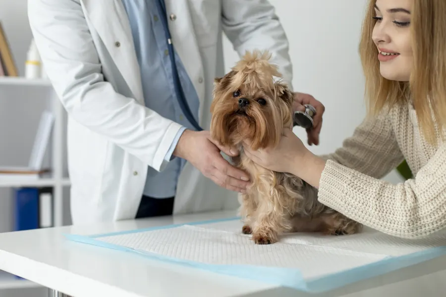
When to see a vet urgently
Pain or disrupted bodily functions associated with an inguinal hernia may indicate an emergency. Emergency assessment is needed if:
- A groin swelling becomes hard, markedly swollen or painful.
- Your dog vomits and has abdominal pain.
- Urination becomes difficult or your dog cannot pass urine.
- A female dog develops abnormal vaginal discharge or bleeding.
- Your dog's condition deteriorates rapidly and they cannot stand.
A bladder, bowel or uterus trapped in a hernia requires immediate treatment. You can find a vet near you for help.
Overview
A swelling forms between the abdomen and hindlimb
The inguinal opening is a normal opening in the abdominal wall near the pelvis. If it is too wide, tissue can enter the inguinal canal or pass beneath the skin. A hernia consists of the opening, hernial sac and its contents. Contents may include fat or omentum, the fatty tissue supporting abdominal organs.
A swelling may appear in one groin or both. A hernia containing only fat does not usually cause signs of general illness. It may nevertheless contain organs such as bowel, bladder or uterus. Identifying the contents therefore affects treatment urgency.
Among adult males studied, an inguinal hernia was one-sided in 17 of 20 dogs, or 85%. Of these, 10 were on the left and 7 on the right. Bilateral hernias occurred in 3 of 20 dogs, or 15%. These figures concern acquired, nontraumatic hernias in males.
Causes
A congenital or acquired hernia
A congenital hernia is present from birth. A hernia developing later may involve muscle weakness or an accident. Trauma can damage and weaken the lower abdominal muscles. Other abdominal hernias may occur with the same injury.
A hernia can pass through an existing anatomical opening, such as the inguinal ring. Congenital hernias are usually indirect hernias of this kind. A direct hernia instead passes through a tear in the abdominal wall.
Hormones, tissue strength and inheritance
Oestrogen can weaken collagen in groin connective tissue. The weight of a pregnant uterus or fat associated with obesity can stretch an already weakened inguinal opening. Excess glucocorticoid effects can also weaken the abdominal wall.
An inherited predisposition involving several genes is suspected in Cocker Spaniels and Dachshunds. This differs from a single genetic defect established across every breed.
Risk factors
Acquired inguinal hernias are diagnosed particularly in middle-aged and older, small, unspayed females. A female's inguinal canal is shorter and wider than a male's. Congenital inguinal hernias occur more often in young small males. Breeds described as predisposed include Basenjis, Pekingese, Poodles, Cairn Terriers and West Highland White Terriers.
An acquired inguinal hernia is nevertheless possible in an adult male. The 21 adult males studied had a mean age of 8.7 years and standard deviation of 2.7 years. Median weight was 9.8 kg, ranging from 2.6–49.9 kg. Such a patient group does not define the age or size of every dog with an inguinal hernia.
In the adult male hernia group, 9 dogs were neutered and 12 intact. An acquired inguinal hernia was therefore possible in both groups.
Typical symptoms
An inguinal hernia may be only a local swelling or involve general signs as well. At home, you may notice:
- A soft, usually painless groin swelling.
- A harder, larger, tender swelling if the hernia is complicated.
- Redness or bruising over the swelling.
- Vomiting, diarrhoea or abdominal pain.
- Difficulty urinating.
- Tiredness and being more subdued than usual.
- Abnormal vaginal discharge or bleeding in a female.
An uncomplicated hernia may be reducible into the abdomen during veterinary examination. A trapped, painful mass may not return. Reducibility is a finding your vet assesses alongside the rest of the situation.
Among adult males without a concurrent perineal hernia, 9 of 13 dogs, or 69.2%, had groin swelling as their only reported sign.
Scrotal or testicular swelling alongside groin swelling has also been described in males. Reduced appetite is another possible sign.
Complications and consequences
Organ entrapment and strangulation
An organ entering a hernia can become trapped in the opening, or incarcerated. In a strangulated hernia, the organ's blood supply may also be compromised. A damaged bowel segment may need removal during surgery. Peritonitis is one complication requiring immediate surgery. The outlook for a complicated hernia depends on how much organ damage occurs before repair.
A concurrent perineal hernia in a male
In a group investigating acquired inguinal hernias in males older than 1 year, 8 of 21 dogs, or 38.1%, also had a perineal hernia. A perineal hernia is in a different area and can cause difficulty passing faeces or obstruction associated with bladder displacement. Assessment of an adult male with an inguinal hernia therefore also checks for a perineal hernia. The figure does not concern females or puppies with a congenital inguinal hernia.
Diagnosis at the vet
Examination identifies the swelling's contents
Your vet may feel an enlarged hernial opening in the groin. Softness, tenderness and reducibility are assessed at the same time. Ultrasound or X-rays usually establish whether organs are involved. The swelling's external size alone therefore does not determine its contents.
Even an easily reducible hernia may contain bowel. Ultrasound can show bowel or fat passing through the inguinal canal. An ultrasound-guided fine-needle sample sometimes helps identify the nature of a swelling. CT or MRI may provide additional investigation where needed.
In a reported puppy with bilateral inguinal hernias, preoperative laboratory tests were normal. Ultrasound nevertheless showed that part of the small intestine was also present in the hernias. In the same dog, the contents could not be felt when lying on their back because they readily returned to the abdomen.
Abnormal-looking hernial contents can be sampled for histology during surgery. In one adult male, adenocarcinoma was found in such a fat tissue sample. This individual finding does not mean an ordinary inguinal hernia is a tumour.
Blood tests and general condition
A blood count can detect increased white blood cells, which may accompany bowel strangulation. Biochemical blood tests assess disturbances including electrolyte imbalance. Urine tests may also form part of initial investigations. The combination of pain, vomiting and urinary difficulties affects the urgency of investigation and surgery.
An adult male's perineal area is also assessed before planning surgery. Inguinal and perineal hernias may require separate surgical procedures. This does not mean every dog with an inguinal hernia has both conditions.
Treatment
Corrective surgery and its timing
An inguinal hernia is usually repaired surgically after diagnosis because delay can make the procedure more difficult. A hernia involving pain, peritonitis or organ entrapment requires immediate surgery. The aim is to return tissues to the abdomen and close the abnormal opening. Damaged tissue is removed where necessary. Your vet selects treatment using examination findings and the dog's condition.
Open surgery can use an approach over the groin or through the abdominal midline. The midline approach also allows abdominal exploration. Where possible, the hernia opening is closed using healthy surrounding tissue. Repair takes account of the blood vessels and nerves crossing the groin.
Laparoscopic repair and other procedures
Selected dogs can undergo laparoscopic hernia repair. A described technique was particularly suitable for easily reducible congenital hernias. Adhesions and nonviable tissue can make laparoscopy more difficult. Suitability is therefore not determined by wound size alone.
Among 16 operated adult males, 15 openings were closed by suturing tissues together. One dog required mesh for repair. Neutering can also be performed at the same time if included in the treatment plan.
Monitoring a puppy's hernia
Some congenital inguinal hernias may close by around 12 weeks of age. Your vet assesses whether monitoring is suitable or repair is needed. Organ entrapment or severe pain requires urgent treatment in a puppy as well. Monitoring therefore does not mean waiting at home with a painful hernia.
Recovery and outlook
The surgical prognosis for an uncomplicated inguinal hernia is generally excellent. A strangulated hernia has a more guarded outlook depending on organ damage. In the adult male group, postoperative complications occurred in 3 of 16 dogs, or 18.8%. These included aspiration pneumonia, a scrotal haematoma and constipation.
Follow-up information was available for 14 dogs. None had a recorded recurrence by their last follow-up. Median follow-up was nevertheless only 18.5 days, ranging from 2–1,697 days. The result therefore does not describe each dog's lifelong recurrence risk.
A 7-month-old male puppy treated laparoscopically had groin swellings around 2–4 centimetres in diameter before repair. The dog went home 1 day after surgery. No recurrence was seen at 1 year. This was one dog's outcome and does not determine discharge timing for other patients.
Prevention
Maintaining a healthy weight may reduce acquired inguinal hernia risk. Spaying reduces oestrogen effects and may lower risk associated with tissue weakening. Breeding from a male with a congenital inguinal hernia is not recommended. Some Finnish Lapphund breeding guidelines recommend against breeding from a dog diagnosed with an inguinal hernia.
Prevent licking and scratching of the surgical wound during recovery. A collar provides protection where needed. A dirty or wet dressing is changed.
Breeding restrictions for congenital inguinal hernia reflect the possibility of passing predisposition to offspring.
Important for dog owners
Observations before and after surgery
Record changes in the groin swelling's size, tenderness or skin colour. Monitor vomiting, urination and general condition. A hard, painful hernia and other general signs require prompt veterinary assessment.
After discharge, watch for wound redness, swelling and discharge. Thick or foul-smelling discharge and increasing pain are reasons to contact the treating vet. Prevent licking or scratching of the surgical area. Arrange follow-up and increases in exercise according to discharge instructions.
Frequently asked questions
Is a soft inguinal hernia harmless?
An uncomplicated inguinal hernia is often soft and painless. Even an easily reducible hernia may nevertheless contain bowel. Your vet therefore uses imaging to identify its contents. An absence of pain alone is not a reason to leave a new groin swelling unexamined.
Can a puppy's inguinal hernia disappear on its own?
A congenital hernia may close spontaneously during the first approximately 12 weeks of life. Your vet assesses whether monitoring is appropriate using the puppy's findings. Severe pain or an organ trapped in the hernia requires immediate treatment. This age threshold is therefore not a reason to postpone examining a puppy with signs.
Does an inguinal hernia always need mesh?
Mesh is not part of every repair: in the operated adult male group, 15 dogs had the opening closed by suturing tissues together. One dog's repair required mesh. Surgery aims to use healthy, strong surrounding tissue. The method is tailored to the opening being repaired.
Can a male dog also have an inguinal hernia?
Congenital inguinal hernias occur particularly in young small males. An older male can also develop an acquired inguinal hernia. Examination of an adult male also checks for a possible perineal hernia. A male dog's groin swelling is therefore examined just as a female's is, although the most common patient groups differ.
When should I contact a vet about the surgical wound?
Increasing swelling, redness and pain, and thick or foul-smelling discharge, are reasons to contact your vet. A collar prevents wound licking where needed. Check discharge instructions for wound protection, agreed follow-up visits and which changes to report to the treating vet.
Sources
- Inguinal Hernia (2026), VCA Animal Hospitals, accessed 2026-11-08.
- Evaluation of concurrent perineal hernia in adult male dogs presenting with nontraumatic, acquired inguinal hernias (2021), Canadian Veterinary Journal, accessed 2026-11-08.
- Suomenlapinkoirien jalostuskriteerit: nivustyrä, Lappalaiskoirat ry, accessed 2026-11-08.
- Bilateral intracorporeally sutured inguinal herniorrhaphy using 3-dimensional laparoscopy in a dog (2021), Canadian Veterinary Journal, accessed 2026-11-08.
- Mitä on päivystyshoito, Helsingin yliopisto, accessed 2026-11-08.
- Lemmikin haavanhoito, Eläinlääkäriasema Lempiellit, accessed 2026-11-08.
- Koirien tyrät ja niiden kirurginen hoito, Delivet, accessed 2026-11-08.
- Hernias in Animals (2024), Merck Veterinary Manual, accessed 2026-11-08.
This information does not replace a visit to a vet. In an emergency, contact your nearest emergency vet: find a vet (RCVS).
More on this topic: Bones, joints and muscles
- Atlantoaxial instability in dogs: neck pain and treatment
- Crooked front legs in puppies: angular limb deformity
- Cruciate ligament injury in dogs – a torn cranial cruciate ligament
- Hip dysplasia in dogs: symptoms, tests and treatment
- Immune-mediated polyarthritis in dogs: symptoms and care
- Tendonitis in dogs – symptoms, investigations and treatment