
Dog diseases · Heart and blood vessels
Pericardial effusion in dogs: symptoms and treatment
Pericardial effusion means excess fluid within the sac surrounding the heart. Fluid can compress the heart and reduce blood pumping to the body. Sudden obstruction of cardiac filling, called tamponade, requires immediate veterinary care.
When to see a vet urgently
Circulatory collapse associated with tamponade can rapidly be fatal.
- Seek emergency care if your dog collapses or loses consciousness.
- Breathing difficulty warrants immediate contact with an emergency vet.
- Very pale gums with weakness may indicate impaired circulation.
- Rapid deterioration in general condition is an emergency sign.
- After previous effusion treatment, renewed collapse may indicate recurrence.
Overview
The pericardial sac normally contains only a little lubricating fluid to ease heart movement. Excess fluid increases pressure within it. In tamponade, the chambers cannot fill normally. This reduces the volume of blood pumped by the heart.
Pericardial effusion has been identified in about 7% of dogs investigated for cardiac symptoms. In one northern European hospital series of 82 patients examined in 2008–2018, 79% had findings consistent with acute tamponade. Slowly accumulating fluid may initially cause few symptoms because the sac has time to stretch. Pressure generally compresses the right atrium first, then the right ventricle.
Causes
Tumours and inflammation are the most common causes
The most common canine causes are a cardiac tumour and idiopathic pericarditis. Idiopathic means the actual cause of inflammation cannot be identified. Haemangiosarcoma is a malignant tumour occurring particularly around the right atrium or atrial appendage. When it bleeds into the sac, fluid red blood cell concentration may resemble ordinary blood. A heart-base tumour such as a chemodectoma can also cause effusion.
Among 29 effusion patients examined in 2016–2020, 19 had a tumour-related cause (66%). Idiopathic inflammation explained 10 cases in the same group (34%). Of the 19 dogs with tumours, 12 had haemangiosarcoma (63%).
Other possible causes
Chest trauma or a clotting disorder can cause bleeding into the sac. Rupture of the left atrium can also cause fluid accumulation. Bacterial infection can spread to the pericardium through systemic infection or a migrating foreign body.
A rare cause is low blood albumin, allowing fluid to escape vessels more readily. A congenital diaphragmatic hernia can also be associated with effusion.
Risk factors
Effusion occurs particularly in middle-aged and older dogs. Males and large breeds are overrepresented. Predisposed breeds include Golden Retrievers, Great Danes and Pyrenean Mountain Dogs. Breed risk partly reflects the frequency of underlying diseases.
Dogs with cardiac haemangiosarcoma are typically large and over 7 years old. Idiopathic pericarditis often affects medium or large dogs over 6 years. Heart-base chemodectomas occur particularly in short-muzzled breeds. Pericardial effusion has also been diagnosed in a 6-month-old dog. In the northern European series of 82 patients, 17 weighed under 15 kg.
Typical symptoms
Severity depends strongly on how rapidly fluid accumulates. Early signs of gradual accumulation may include abdominal enlargement and reduced exercise tolerance.
- The abdomen swells because of fluid accumulation.
- Your dog tires on shorter walks than usual.
- Breathing becomes faster or laboured.
- Appetite decreases.
- Your dog may vomit.
- Exertion can cause fainting.
- Your dog may also cough.
Longstanding milder disease can cause muscle loss. Infection-associated effusion may cause fever too. Severe, rapidly developing effusion can cause collapse without previous warning signs. When a tumour ruptures, serious circulatory impairment may occur before abdominal swelling has time to develop.
Complications and consequences
Impaired circulation and recurrence
Cardiac compression raises venous pressure and causes right-sided heart failure signs. Tissues then receive insufficient oxygen. In tamponade-related shock, mechanical compression restricting cardiac filling obstructs circulation.
Persistent pericarditis can rarely scar and stiffen the sac. A stiffened sac can restrict heart function without a large fluid volume. Idiopathic effusion recurs in about 50% of patients.
Outlook depends on the cause
Non-tumour-related effusions generally respond well to appropriate treatment. For idiopathic inflammation, expected survival after pericardial surgery is about 600 to over 1,000 days. Right atrial haemangiosarcoma has a poor outlook because it has often spread by diagnosis. After surgery and chemotherapy for haemangiosarcoma, survival may be 3–6 months. With a heart-base tumour-related effusion, a dog may live up to 2 years after successful surgery. Survival after different chemodectoma treatments has ranged from 6 months to 3 years. Without additional treatment, haemangiosarcoma-associated fluid often reaccumulates within 1 day to 3 weeks.
Diagnosis at the vet
Ultrasound confirms fluid and cardiac compression
Your vet first assesses breathing and circulatory adequacy. Muffled heart sounds, a weak pulse and rapid heart rate raise suspicion. Neck veins may become distended if blood backs up on the right side. Low blood pressure also supports suspected tamponade.
Heart ultrasound is the most sensitive and accurate method of demonstrating effusion. It shows whether the right atrial or ventricular walls collapse. Emergency focused ultrasound helps identify an urgent need for drainage. A fuller cardiac examination then investigates a possible tumour.
Other tests investigate the cause
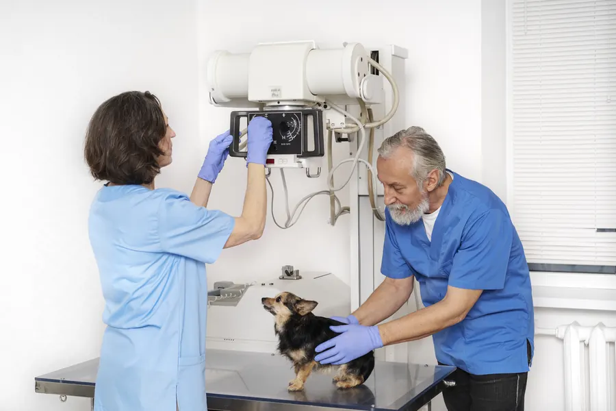
On X-rays, the cardiac silhouette may be larger and rounder than usual. However, a normal silhouette does not exclude effusion. ECG shows rhythm and arrhythmias. Heart movement within fluid may cause alternating ECG wave heights.
Blood tests assess general health and possible other diseases. Routine blood and urine results can be normal despite effusion. Removed fluid can be assessed for cells and inflammation. Cytology is particularly helpful for identifying infection or lymphoma. Among fluid samples from 259 dogs, cytology established an underlying diagnosis in only 20 cases (7.7%). When fluid packed cell volume was below 10%, cytology identified the cause in 20.3%. Fluid pH poorly distinguishes tumour-related from other effusions.
Bloody fluid alone cannot distinguish cancer from idiopathic inflammation. If needed, chest imaging and abdominal ultrasound assess tumour spread. Small haemangiosarcoma metastases may not appear on initial chest X-rays. Surgical tissue samples can distinguish pericardial mesothelioma from idiopathic inflammation. An ultrasound-invisible tumour remains possible even without an obvious mass.
Treatment
Acute tamponade
In tamponade, your vet drains fluid by pericardiocentesis. A catheter is introduced into the sac and fluid is withdrawn through it. Ultrasound can help select a safe insertion site. Sedation and local anaesthesia may be used. ECG monitors rhythm during the procedure.
A catheter touching heart muscle can trigger an arrhythmia. Afterwards, blood pressure and rhythm recovery are monitored. Serious complications are described as uncommon, although estimates of 10–15% have also been reported. Intravenous fluids may support circulation. In a 30-dog comparison, fluids before drainage did not measurably improve circulatory status assessed by the heart rate to systolic pressure ratio.
Diuretics can worsen acute tamponade by reducing circulating fluid volume. Routine medication generally cannot resolve the problem caused by fluid within the sac. A small effusion in a stable patient can sometimes be investigated further before drainage.
Recurrent effusion and pericardial surgery
Drainage relieves acute compression, but the cause needs a separate treatment plan. Idiopathic effusion sometimes settles after a single drainage procedure. If fluid returns, the procedure may need repeating. Recurrent cases may undergo surgery to remove part of the sac.
Surgery creates a route for fluid to leave the area around the heart, preventing pressure accumulating in the same way. A dog can live without the pericardium. Surgery can be performed through an open chest approach or thoracoscopy. The heart can also be inspected for tumours missed on ultrasound.
Options for a tumour patient may include surgery, chemotherapy, radiotherapy or symptom relief. Infectious pericarditis needs treatment targeting its cause.
Recovery and follow-up
Dogs often go home within 2–5 days after pericardial surgery. Initially, keep your dog very quiet until stitches are removed, usually after 10–12 days. Exercise remains restricted for another 3–4 weeks before a gradual return to normal walks. A dressing protects the surgical area until stitch removal.
For recurrent or tumour-associated effusion, ultrasound checks may occur every 3–4 months. New signs suggesting tamponade require examination before the scheduled check. Continued postoperative bleeding can cause chest cavity inflammation and breathing difficulty.
Prevention
No specific diet or supplement has been shown to prevent pericardial effusion. The cause of idiopathic inflammation remains unknown. Corticosteroids have not been shown to prevent its recurrence. Treating confirmed infection or other underlying disease forms part of controlling further accumulation.
Monitoring after drainage helps identify recurrent symptoms. Surgery for repeated effusion can prevent renewed compression. Tumour patients continue regular ultrasound monitoring after treatment too.
Important for dog owners
Record stamina, appetite, breathing and abdominal size changes. Monitor muscle condition too. Contact your practice immediately if weakness, fainting or breathing difficulty returns. Returning symptoms may require repeat ultrasound and drainage.
After surgery, keep the chest dressing clean and dry. Quiet recovery continues after discharge. Arrange follow-up according to the cause identified. Successful drainage alone does not remove the need to investigate underlying disease.
Frequently asked questions
Does fluid around the heart always mean cancer?
A tumour is a common cause, but idiopathic inflammation can also cause effusion. Other possibilities include trauma and clotting disorders. The sac can contain blood without cancer too. The cause is investigated with heart ultrasound and fluid samples or tissue examination if needed.
Can medicine remove pericardial fluid?
In acute tamponade, accumulated fluid usually needs catheter drainage. A diuretic can worsen circulation by reducing blood volume. Other medication for the underlying disease is selected separately once the cause and circulatory state are established.
Can fluid return after drainage?
Idiopathic effusion recurs in about 50% of dogs. Drainage can then be repeated, and partial sac removal may be considered for frequent recurrence. Renewed weakness or breathing difficulty warrants prompt contact with your vet.
How long can a dog live with pericardial effusion?
The outlook depends crucially on whether inflammation or a tumour is responsible. In idiopathic inflammation, expected survival after surgery is about 600 to over 1,000 days. Right atrial haemangiosarcoma has a substantially poorer outlook. Tumour type influences treatment effectiveness and disease progression.
Can a dog live without the pericardium?
A dog can live without it. The sac has a relatively minor role in the body. Partial removal prevents new fluid compressing the heart. Surgery can suit recurrent idiopathic effusion, for example, but the decision depends on the cause and your dog's circumstances.
Sources
- Sydänpussin nestekertymä koiralla – kirjallisuuskatsaus ja kartoitus tapauksista Yliopistollisessa eläinsairaalassa 2008–2018, Suomen Eläinlääkärilehti, accessed 2026-11-07.
- Pericardial Disease in Dogs and Cats, Merck Veterinary Manual, accessed 2026-11-07.
- Pericardiectomy, North Downs Specialist Referrals, accessed 2026-11-07.
- Acquired Heart and Blood Vessel Disorders in Dogs, Merck Veterinary Manual, accessed 2026-11-07.
- Mitä on päivystyshoito, Helsingin yliopisto, accessed 2026-11-07.
- Retrospective Evaluation of the Causes and Fluid Characteristics of Cavitary Effusions in Dogs and Cats, Journal of Veterinary Internal Medicine, accessed 2026-11-07.
- Basic triage in dogs and cats: Part II, Canadian Veterinary Journal, accessed 2026-11-07.
- Pericardial Effusion in Dogs, VCA Animal Hospitals, accessed 2026-11-07.
- Pericardial Effusion, VetCardio, accessed 2026-11-07.
- Diagnostic Yield of Cytologic Analysis of Pericardial Effusion in Dogs, Journal of Veterinary Internal Medicine, accessed 2026-11-07.
- Heart Disease – Dogs: Pericardial Effusion, Cummings School of Veterinary Medicine, Tufts University, accessed 2026-11-07.
- Intravenous Bolus Fluid Therapy Versus No Fluid Therapy Prior to Pericardiocentesis in Dogs: A Randomized Controlled Trial in 30 Dogs, Journal of Veterinary Emergency and Critical Care, accessed 2026-11-07.
This information does not replace a visit to a vet. In an emergency, contact your nearest emergency vet: find a vet (RCVS).
More on this topic: Heart and blood vessels
- Boxer cardiomyopathy (ARVC) in dogs
- Dilated cardiomyopathy in dogs: symptoms, tests and treatment
- Mitral valve disease in dogs: symptoms, tests and treatment
- Arrhythmias in dogs: symptoms and treatment
- Sick sinus syndrome in dogs – symptoms and pacemaker treatment
- Heart failure in dogs: symptoms, tests and treatment