
Dog diseases · Urinary system
Urinary blockage in male dogs – symptoms and emergency treatment
A urethral blockage prevents urine flowing out of the bladder. It is a life-threatening emergency for your dog. A male's narrow urethra particularly predisposes to stones becoming lodged. If your dog strains without passing urine, they need a vet immediately. Even a partial blockage may cause only drops or a weak stream.
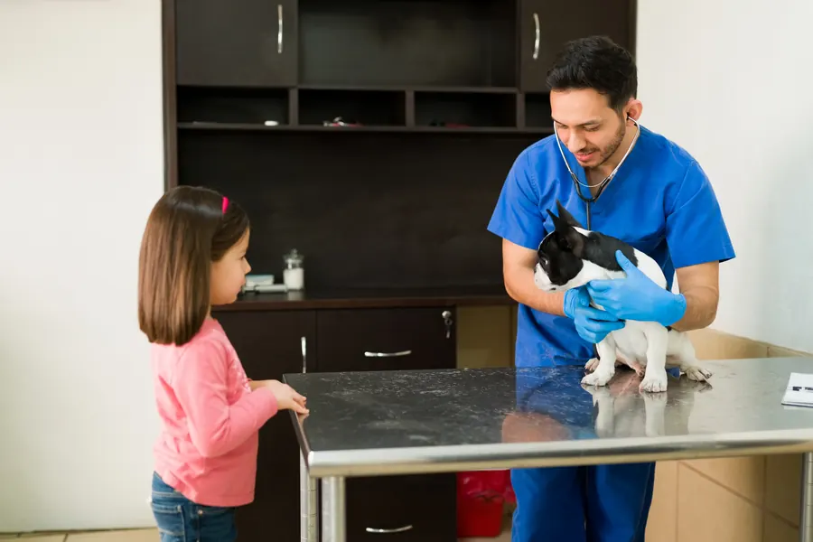
When to see a vet urgently
Do not monitor inability to urinate at home until the next day. Seek emergency care particularly if:
- Your dog adopts a urination posture and strains but passes no urine.
- Attempts to urinate are painful and your dog vocalises.
- Your dog becomes quiet, refuses food or starts vomiting.
- The abdomen is painful or swells.
- Your dog collapses or becomes less conscious.
A complete blockage can cause uraemia within 36–48 hours. Untreated obstruction can result in death in around 72 hours. Do not wait for these times to pass before seeking help. You can find a vet near you.
Overview
Why does a male dog's urethra become blocked?
A male's urethra runs through the rear of the body, between the hindlegs and into the penis.
The urethra carries urine from the bladder out of the body. In the male's penis, it runs in a groove in the penile bone and narrows. Stones can lodge behind the penile bone. Urinary stones are the most common cause of urethral blockage in dogs.
In one study from northern Europe examining 2,306 urinary stone samples from 1982–2012, 52% contained calcium oxalate. Struvite stones accounted for 37%. Ammonium urate stones accounted for 5.4%. Cystine stones accounted for 3.1%. These figures describe the composition of stones examined during that period, rather than the current blockage risk for all dogs.
Causes
A stone, narrowing or another obstruction
A stone entering the urethra creates a physical barrier to urine. A urinary tract tumour can also close the passage. Scarring after injury or a previous procedure can narrow the urethra. A fractured penile bone is another possible cause of blockage in males.
Obstruction can also result from pressure outside the urethra. Prostatitis, an abscess or a cyst can contribute to mechanical urinary obstruction. Blood clots, foreign objects and inflammatory tissue changes are other possible causes.
A blockage raises pressure in the bladder and the urethral segment between the bladder and obstruction. Urinary stones form when minerals in urine precipitate under suitable conditions, first as crystals and then as stones.
A functional disorder can also prevent urination without an object wedged in the urethra. Coordination between bladder and urethral muscles is then impaired. Tests distinguish this from a mechanical blockage.
Risk factors
A male's longer, narrower urethra increases the risk of stones becoming lodged. Previous stone formation increases the possibility of recurrent obstruction. Calcium oxalate stones occur more frequently in breeds such as Miniature Schnauzers, Bichons Frises and Shih Tzus.
Dalmatians have an inherited tendency to urate stones. A liver shunt can predispose to ammonium urate stones. Urinary tract infection can promote struvite stone formation. Cystine stones are found particularly in younger entire males. Breed and age help assess the likely stone type, but tests always establish the cause of blockage.
Calcium oxalate stone formation is more common in middle-aged and older dogs, although young dogs can be affected too. In one stone study from northern Europe, calcium oxalate-containing stones were found more often in males than females.
Typical symptoms
With a partial blockage, some urine may still pass despite a clearly altered flow. Possible observations include:
- Repeated attempts to urinate, passing only a little each time.
- Staying in a urination posture longer than usual.
- A weak urine stream or urine passed in drops.
- Visible blood in urine.
- Urinating indoors or on the dog's own bed.
- Straining or showing pain during urination.
With complete obstruction, your dog cannot pass urine despite straining. Tiredness, loss of appetite and vomiting can follow deteriorating general condition. Your vet may find a large bladder that is painful to touch. Signs at home cannot establish whether the obstruction is a stone, narrowing or another change.
Complications and consequences
The kidneys, heart and bladder
Urinary obstruction prevents normal waste elimination. Temporary or permanent kidney dysfunction can result. Blood potassium may rise. High potassium can cause life-threatening heart rhythm disturbances. The body's acid–base balance can also be disrupted.
An overfull bladder can rupture, allowing urine into the abdominal cavity. Obstruction can damage the bladder lining and the detrusor muscle that contracts to expel urine. Chronic blockage can reduce bladder contraction even after the obstruction is relieved.
After surgery to open the bladder, urine can leak through the bladder suture line into the abdominal cavity. A hernia can also develop at the abdominal incision. Surgical site infection is another possible complication.
The blockage and procedures can cause urethral scarring that predisposes to another blockage. The outlook after surgery for stone obstruction can be excellent if prolonged obstruction has not caused severe kidney or systemic damage. The outlook for tumour-related obstruction or extensive scar narrowing is assessed separately according to the underlying disease.
Diagnosis at the vet
Assessment starts with general condition
Your vet assesses bladder filling, pain, dehydration and circulation. Blood tests check kidney values, electrolytes and acid–base balance. An ECG is particularly needed with dangerously high potassium. Test order is adapted to the patient's stability so relief of the blockage is not delayed.
Difficulty passing a catheter can suggest mechanical obstruction. With partial obstruction, a catheter may pass alongside stones. Examination also includes checking the penis and urethral opening for injury, blood or stone material. The urethra and prostate can be felt through the rectum.
Imaging and samples
X-rays look for stones in the bladder or urethra. However, not all stones appear on plain X-rays. Contrast imaging can reveal narrowing caused by a tumour or scar. Ultrasound can investigate bladder tumours, blood clots and stones that are poorly visible on X-rays. The kidneys, ureters and prostate are also assessed if needed.
A urine sample is checked for crystals and signs of infection. It can be sent for bacterial culture. Removed stones are analysed to establish their precise composition. Abnormal bladder tissue may be biopsied. Endoscopy can help assess the inner urethral surface and treat an obstruction.
With calcium oxalate stones, treatable metabolic diseases are investigated, such as high blood calcium, Cushing's disease or hyperparathyroidism. Concurrent kidney or heart disease can also affect treatment and prevention choices.
Treatment
Emergency care and relieving the blockage
Your vet stabilises an unwell patient and treats dangerous electrolyte disturbances. Intravenous fluids are needed to correct dehydration or inadequate circulation, for example. Urine outflow must be restored. Initially, a catheter may be used to try to flush an obstructing stone back into the bladder. This is performed at the practice; do not try pressing the abdomen at home.
If your dog is too unstable for the sedation or anaesthesia needed to relieve the blockage, your vet may drain the bladder with a needle. Emergency measures counteracting potassium's effects are temporary until urine flow and potassium elimination can be restored.
Definitive surgery in a very ill dog may be postponed for 1 or 2 days while catheter drainage and stabilisation take place. Urine already has a drainage route in this situation; your dog is not left waiting with a closed obstruction. Bladder stones can be removed by opening the bladder in a cystotomy under general anaesthesia. In some cases, a stone can be removed endoscopically or broken up with a laser.
Stone type determines further treatment
Struvite stones can be dissolved with a veterinary-prescribed diet only once urine outflow is secure. Complete dissolution takes an average of 1–2 months. Calcium oxalate stones do not dissolve with medication or diet and must be removed mechanically. Treating infection-associated struvite disease includes treating the bacterial infection.
When the urethra needs a new outlet
If a blockage cannot be relieved or stones cause repeated problems, a permanent urethral opening can be created in the scrotal area. Urine then exits from the wider urethral section behind the penile bone. An entire dog is castrated during the same procedure. Large stones can still block the bladder or upper urethra.
The penis is usually left in place during this urethrostomy, but urine passes through the new opening. With a penile tumour, the penis may also need removal.
In some situations, a stent can hold open an obstruction caused by a tumour or narrowing. Relief of obstruction has been reported in 97.6% of tumour patients receiving stents. Urinary incontinence after stenting has been described in 26–41%. These figures concern selected stent patients, rather than surgical outcomes for all stone blockages.
Care after surgery
Skin stitches after urethrostomy are removed after 10–14 days if needed. Wound licking or marked excitement can increase bleeding from the new opening.
After bladder surgery, your dog may need to urinate more often for a few weeks. Urine may contain a little blood. Clear active bleeding requires contacting the treating practice. A protective collar is usually worn for 10–14 days to protect the wound. Possible problems after urethrostomy include bleeding, urine irritation of the skin, scarring of the opening and infection. Follow-up assesses the urine stream, residual urine and recovery of blood values.
Prevention
Recurrent stone blockage is prevented by reducing factors involved in forming that particular stone type. Moist food and good water intake reduce urine concentration. Your dog should have several opportunities to empty the bladder during the day.
With frequently recurring calcium oxalate stones, X-rays every 3–6 months may detect new stones while small. After infection-induced struvite disease, investigating the infection's cause and monitoring recurrence are particularly important. A long-term special diet is not automatically needed once the infection has resolved. Castration may be recommended for a male forming cystine stones to prevent androgen-dependent recurrence.
A low-purine diet may be used to prevent urate stones. Cystine stones often recur within 1 year unless preventive measures are taken. High blood calcium predisposing to calcium oxalate stones is corrected where possible.
Important for dog owners
Watch the urine stream and follow the individual care plan
Record the last normal urination, onset of signs, previous stones and urinary tract infections. Bring a medication list and details of your dog's current diet. After discharge, watch whether urine passes in a continuous stream or only drops, and whether urination looks painful. Observing the urine stream forms part of monitoring after obstruction.
Use the protective collar and restrict exercise as instructed by the treating practice. Ask for the stone analysis result and how it affects prevention, as further care differs between stone types. Arrange follow-up samples and imaging before discharge. Even a urethrostomy does not eliminate every possibility of future blockage. If urination stops again or your dog's condition deteriorates, contact an emergency vet immediately. Do not wait for a scheduled check.
Frequently asked questions
Can a dog have a blockage while passing a little urine?
Yes. With partial obstruction, your dog may pass small amounts frequently or only drops. A few drops therefore do not show that the urethra is properly open. Pain, repeated attempts and a weakening stream warrant investigation without delay.
Why do stones particularly lodge in a male dog's urethra?
A male's urethra is longer and narrower than a female's. It narrows further at the penile bone, and a stone can lodge behind it. A stone formed in the bladder can therefore cause sudden outflow obstruction as it travels with urine towards the outside of the body.
Can a blockage be treated with a dissolution diet?
A complete urethral blockage is not treated by waiting for stones to dissolve. Your vet first establishes urine drainage. Dissolving struvite stones safely within the bladder can take an average of 1–2 months. Calcium oxalate stones cannot be dissolved with diet or medication and require mechanical removal.
Does every blockage need a urethrostomy?
No. Some blockages can be relieved with a catheter and stones moved back into the bladder for removal. A permanent urethral opening is considered for situations such as an unrelievable blockage or recurrent stone problems. The method depends on the cause of obstruction and urethral condition, and further care is planned after surgery too.
Can a dog develop another blockage after surgery?
Yes. A scar developing in the urethra can cause new narrowing. After urethrostomy, large stones can still cause obstruction higher in the urinary tract. Prevention and monitoring tailored to stone composition therefore continue even after successful relief.
Sources
- Urinary Obstruction in Dogs, American College of Veterinary Surgeons, accessed 2026-11-08.
- Urethral Obstruction in Small Animals, Merck Veterinary Manual, accessed 2026-11-08.
- ACVIM consensus statement on diagnosis and management of urinary incontinence in dogs (2024), Journal of Veterinary Internal Medicine / ACVIM, accessed 2026-11-08.
- Mitä on päivystyshoito, Helsingin yliopisto, accessed 2026-11-08.
- Virtsakivet koiralla – oireet ja hoito, FirstVet, accessed 2026-11-08.
- Disorders of Micturition in Dogs and Cats, Merck Veterinary Manual, accessed 2026-11-08.
- Urolithiasis in Dogs, MSD Veterinary Manual, accessed 2026-11-08.
- Virtsakivien koostumus suomalaisilla koirilla 1982–2012 (2018), Suomen Eläinlääkärilehti, accessed 2026-11-08.
- Eläinlääkärin palvelut koirille – virtsateiden sairaudet, Paras Kaveri, accessed 2026-11-08.
This information does not replace a visit to a vet. In an emergency, contact your nearest emergency vet: find a vet (RCVS).
More on this topic: Urinary system
- Acute kidney injury in dogs – sudden kidney failure
- Bladder stones in dogs: symptoms, tests and treatment
- Ectopic ureter in dogs: urine leakage and treatment
- Chronic kidney disease in dogs
- Kidney infection in dogs – pyelonephritis symptoms and treatment
- Poisoning and kidney damage in dogs – dangerous substances