
Dog diseases · Reproductive system
Vaginitis in dogs: discharge, symptoms and treatment
Vaginitis may cause mucus-like discharge and licking around a dog's vulva. Mild inflammation in a young bitch often resolves after her first season. In adults, an underlying problem usually predisposes to inflammation.
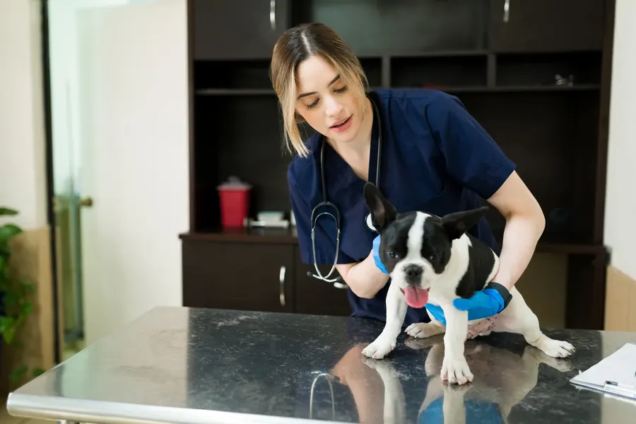
When to see a vet urgently
See a vet immediately if discharge or irritation around the vulva is accompanied by any of the following:
- Your dog becomes noticeably lethargic and loses its usual appetite.
- An unspayed bitch starts drinking much more, particularly after a season.
- The abdomen swells or your dog vomits and seems unwell.
- Your dog collapses or suddenly deteriorates.
These signs may indicate pyometra, an infected womb requiring urgent care. You can find a vet near you for help.
Overview
Vaginitis means inflammation of the vagina or its vestibule. The vestibule is the area between the vulva and the vagina itself. The urinary and reproductive tracts open into the same vestibule.
The vagina extends from the vestibule to the cervix. Its average length in a medium-sized bitch is about 10 cm. Inflammation may have a non-infectious cause.
Juvenile vaginitis occurs before sexual maturity, usually in a bitch under 1 year old. It may be noticed incidentally during a routine health check. Adult-onset vaginitis begins after the first season.
Causes
The exact cause in young dogs often remains unidentified. Immaturity of the reproductive and immune systems may predispose a puppy to inflammation. In adults, urinary incontinence can leave urine irritating the vaginal area.
Vaginal narrowing may prevent normal drainage of urine and secretions. An abnormally positioned ureter is one possible structural problem predisposing to inflammation. Faecal contamination around the vulva may also be involved.
A foreign body in the vagina can cause local irritation. Mucosal injury during mating is another possible cause. A tumour may interfere with drainage from the same area.
Inflammation may involve overgrowth of bacteria such as Escherichia coli or streptococci. Canine herpesvirus can cause genital inflammation. Brucella canis is a rare but relevant infectious cause of vaginitis.
Chemical irritation can also trigger vaginal mucosal inflammation. A vaginal haematoma may underlie inflammation after injury, for example. A local abscess, or collection of pus, is another possible source.
Vaginitis may also occur with uterine infection after giving birth or an abscess in the uterine stump.
Risk factors
Vaginitis can affect bitches of any age or breed. Both spayed and unspayed dogs can develop it. Adult-onset vaginitis is commonly found in spayed bitches.
A vulva hidden by skin folds may predispose to vaginitis. This structural problem is not confined to one breed. A recessed vulva is more common in spayed medium, large and giant dogs.
Excess weight can worsen problems caused by folds covering the vulva. Urine and secretions collecting in the folds create a moist environment for microbes. A concurrent urinary tract infection is one possible underlying factor.
Typical symptoms
Signs you may notice at home include:
- Mucus or pus-like discharge from the vulva appears on the coat or bedding.
- Your dog licks the area more than usual and pays it constant attention.
- Your dog asks to go out or squats to urinate more often during the day.
- Your dog rubs or scoots its bottom along the floor to ease irritation.
- The vulval area looks red or swollen rather than normal.
- Male dogs may show interest even when the bitch is not in season.
In puppies, discharge may be scant, clear or cloudy and sticky. General symptoms are rare in ordinary juvenile vaginitis. Blood in the discharge is less common than mucus or pus-like discharge.
Complications and consequences
The problem may recur if a predisposing structural abnormality is not corrected. Inflammation associated with an uncorrected genital abnormality may ascend to the uterus. Such persistent reproductive tract inflammation can reduce fertility.
Many dogs' symptoms settle within 2–3 weeks of starting treatment. In persistent or structurally related vaginitis, the outlook depends on problem severity and duration. Brucella canis infection can cause miscarriages and puppy deaths.
Genital herpesvirus infection may involve early embryonic death and foetal resorption. Brucella canis can also pass from dogs to people. However, transmission from a symptom-free dog to an otherwise healthy person is very unlikely.
Diagnosis at the vet
Assessment begins with the history and clinical findings. The vet checks vulval structure and surrounding skin folds. Manual vaginal examination may reveal narrowing, an abnormal membrane or a tumour.
Urine sampling and cell examination
Bladder infection is investigated because it may occur at the same time. Urine may be collected by a needle directly into the bladder, called cystocentesis. Some national guidance for acute canine urinary tract infection recommends culture and susceptibility testing of a cystocentesis sample.
A vaginal cell sample is examined for inflammatory and other abnormal cells. The stage of the reproductive cycle is considered during interpretation. The phase after a season can normally involve many neutrophil white blood cells.
Bacterial growth in a vaginal sample from a symptom-free dog does not by itself mean infection. Bacteria in a dog with vaginitis may resemble the normal vaginal microbiota. Susceptibility testing helps select an appropriate antibiotic when medication is needed.
Routine blood and urine tests may be normal in a dog with vaginitis. Cytology or skin scraping of an inflamed fold may add to examination of the vulval area. Blood counts and biochemistry can look for signs of concurrent disease or kidney problems.
Neutrophils may show signs of breakdown in bacterial inflammation. Cell samples from chronic inflammation may contain more lymphocytes and macrophages.
Brucella canis is considered particularly if discharge accompanies miscarriages or other reproductive problems. Antibody testing is the main screening method for symptom-free brucellosis. Symptomatic infection can be confirmed, for example, by bacterial culture of blood or uterine discharge.
Vaginoscopy and further investigations
Vaginoscopy examines the vaginal lining with a camera. It can locate a foreign body, tumour or mucosal ulcer. Painful inflammation may require sedation for examination.
The examination also establishes whether discharge comes from the uterus, vagina or vestibule. The narrow front part of the vagina is about 5–7 mm in diameter. The instrument must therefore be small enough for the dog examined.
Ultrasound or contrast X-rays can help investigate structural abnormalities. If pyometra is suspected, uterine ultrasound helps confirm the diagnosis. Biopsy of suspicious tissue can distinguish inflammation from a tumour.
In a northern European lower urinary tract endoscopy series, the vagina was examined in 43 bitches. Vaginal abnormalities were seen in 8 of them. Vaginal biopsies identified inflammation and a benign inflammatory polyp.
Treatment
The vet chooses treatment according to the cause identified. Mild vaginitis does not always need medication. Keep the vulval area clean in a mildly affected dog to help prevent secondary bacterial infections.
Monitoring young dogs and timing spaying
Juvenile vaginitis may resolve on its own after the first season. If vaginitis is diagnosed before planned spaying, surgery is postponed until inflammation has settled. If symptoms stop before the first season, there is no need to wait for that season before spaying.
Treating the underlying cause
Narrowing, urinary incontinence and possible pyometra are investigated or treated before antibiotics. Any antibiotic needed is chosen according to culture and susceptibility results. Antibiotics can also alter the normal vaginal bacteria.
A mild cleanser recommended by the vet may be used externally. Internal vaginal flushing is not generally recommended. Removing a foreign body or tumour may require surgery.
Corrective surgery for skin folds
Skin folds covering the vulva can be removed when needed by vulvoplasty, also called episioplasty. Surgery is considered particularly when a severe structural problem is thought to trap urine. Active skin inflammation in the area is treated beforehand.
Before anaesthesia, blood tests can assess red cells, white cells and platelets. Abnormal results may need further investigation or treatment before planned corrective surgery.
Correction is performed under general anaesthesia. Excess skin and, when needed, underlying fat are removed. Closing the wound lifts the skin fold, allowing the vulval area to dry more effectively.
Non-dissolving skin stitches are usually removed after 10–14 days. Exercise is restricted for the first 2 weeks after correction. A protective collar is usually used for 1–2 weeks to prevent licking.
Wound breakdown is a possible complication. If too much excess skin remains, the original symptoms may continue. Urinary incontinence may persist despite successful vulval correction.
Pain and swelling after skin fold correction may be treated with a non-steroidal anti-inflammatory drug prescribed by the vet. Excessive running and jumping can reopen the healing wound. A small proportion of dogs need another operation if the first correction is insufficient.
Prevention
Keeping a mildly affected dog's vulval area clean reduces secondary bacterial infection risk. Weight loss in an overweight dog may ease problems from covering skin folds. Maintaining a normal weight after correction helps prevent new excess folds.
Preventing recurrent urinary tract infections relies on identifying their underlying cause. Some national brucellosis prevention guidance particularly recommends testing imported dogs and dogs from abroad used for breeding.
Unnecessary preventive antibiotics can reduce the protective mucosal microbiota. Repeated or prolonged antibiotic use can also favour infections caused by resistant bacteria.
Important for dog owners
Record when discharge began, its colour, amount and smell, and whether it is constant or intermittent. Note the most recent season and any spaying date. Tell the vet about mating, litters, previous urinary tract infections and all medicines used.
Watch how often your dog urinates, whether it wets its bedding and whether water intake has changed. Photograph visible discharge for the appointment. Ask where the discharge is thought to originate and whether separate urine or vaginal samples are needed. Agree how mild juvenile disease will be monitored and when it should be reassessed.
Absence of discharge alone does not rule out closed pyometra. Pyometra symptoms most often begin 4–8 weeks after a season.
Frequently asked questions
Does vaginitis in a puppy resolve by itself?
Mild juvenile vaginitis often resolves after the first season. Mild inflammation does not always need medication. General symptoms are rare in ordinary juvenile disease. Keep the area clean so local irritation does not predispose to further bacterial infections.
Does vaginitis always need antibiotics?
Antibiotics are not automatic treatment for a mildly affected dog. Bacterial growth in a symptom-free dog's vagina may be part of the normal microbiota. If medication is needed, culture and susceptibility testing guide selection. Possible contributions from urinary incontinence or vaginal narrowing are investigated too.
Can a spayed dog have vaginitis?
Yes, adult-onset vaginitis is commonly found in spayed bitches. Urinary incontinence or a vulva covered by skin folds may underlie it. Spaying alone therefore does not remove every local factor predisposing to vaginal inflammation. Concurrent bladder infection is investigated as part of the assessment.
How does vaginitis differ from pyometra?
Vaginitis affects the vagina or vestibule. Pyometra affects the uterus. Pyometra requires urgent veterinary care. It may cause increased drinking, vomiting and marked tiredness. In closed pyometra, there may be no visible discharge at all.
Why does vaginitis recur after treatment?
Recurrence is common if the original predisposing problem remains uncorrected. Narrowing or urinary incontinence can maintain the build-up of urine and secretions. Treatment then also targets the underlying cause identified. A severe skin fold covering the vulva can be corrected surgically when needed.
Sources
- Vaginitis in Small Animals, Merck Veterinary Manual, accessed 2026-11-05.
- Vaginitis in Dogs, VCA Animal Hospitals, accessed 2026-11-05.
- Recessed Vulva in Dogs, Merck Veterinary Manual, accessed 2026-11-05.
- Vulvar Fold Dermatitis, American College of Veterinary Surgeons, accessed 2026-11-05.
- Vulvoplasty (Episioplasty) in Dogs, VCA Animal Hospitals, accessed 2026-11-05.
- Koiran akuutti komplisoitumaton virtsatietulehdus, Ruokavirasto, accessed 2026-11-05.
- Virtsarakon tähystystutkimukset koirilla Yliopistollisessa pieneläinsairaalassa vuosina 2006–2016, Suomen Eläinlääkärilehti, accessed 2026-11-05.
- Pyometra (infected womb) in dogs, PDSA, accessed 2026-11-05.
- Clinical approach to canine vaginitis, Semina: Ciências Agrárias, accessed 2026-11-05.
- Koiran bruselloosi, Ruokavirasto, accessed 2026-11-05.
This information does not replace a visit to a vet. In an emergency, contact your nearest emergency vet: find a vet (RCVS).
More on this topic: Reproductive system
- Vaginal hyperplasia in dogs: swelling and treatment
- Balanoposthitis in dogs – discharge and inflammation
- Enlarged prostate in dogs – symptoms and treatment
- Prostatitis in dogs: prostate infection and treatment
- Infertility in dogs – causes, investigations and treatment
- Miscarriage in dogs: signs, causes and treatment