
Dog diseases · General conditions
Ascites in dogs: abdominal fluid causes and treatment
Ascites means fluid accumulating in the abdominal cavity. It results from different diseases rather than being one independent disease. Causes include heart or liver disease, low blood protein, inflammation or a tumour. Sudden abdominal swelling, weakness or breathing difficulty requires urgent assessment.
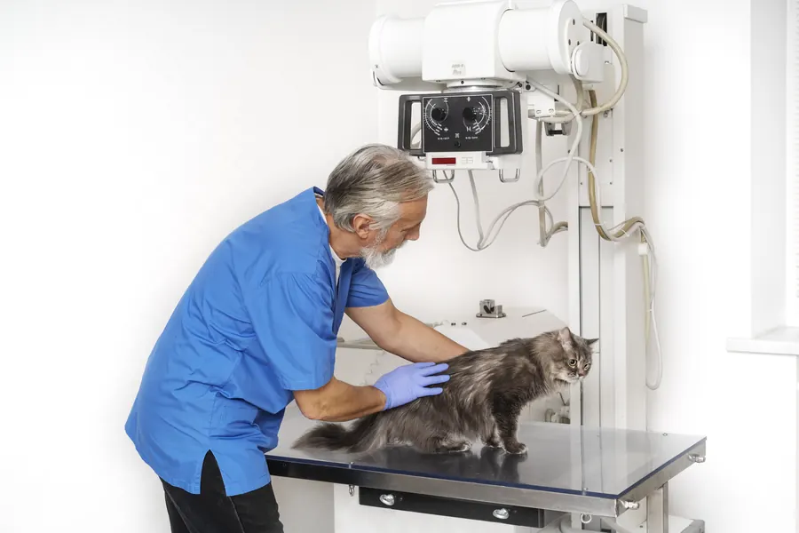
When to see a vet urgently
Seek emergency care if abdominal enlargement accompanies the following signs.
- Breathing is difficult at rest or your dog cannot find a comfortable resting position.
- Your dog collapses, cannot stand or becomes less conscious.
- The mucous membranes are pale or your dog deteriorates rapidly.
- The abdomen is severely painful or swells suddenly.
- Abdominal signs accompany difficulty urinating or vomiting blood.
Cardiac tamponade from pericardial fluid can cause ascites and life-threatening circulatory impairment. Find a vet near you.
Overview
Abdominal fluid may be fluid leaking from the circulation, inflammatory fluid, blood, urine or bile. Treatment therefore does not always mean diuretics alone. Fluid can increase gradually and become visible only once the accumulation is larger. The abdomen may look rounded and pendulous.
However, a bulging abdomen does not confirm fluid, because enlargement has several causes. Imaging helps demonstrate free fluid. Sample characteristics then guide investigation of the cause. Drainage alone may improve comfort, although the underlying disease requires separate treatment.
Causes
Pressure and low protein
Right-sided heart failure can impair blood return and increase abdominal fluid. Obstructed flow within the liver or surrounding vessels can raise portal pressure. Liver fibrosis alters circulatory resistance and can cause portal hypertension. Pericardial pressure can restrict filling of the right side of the heart.
Reduced albumin weakens the blood's ability to retain fluid within vessels. Protein can be lost through the intestine or kidneys. Advanced liver disease may reduce albumin production. Low albumin and raised vascular pressure may both contribute simultaneously.
Prolonged portal hypertension can cause acquired bypass vessels to form. Blood then bypasses the liver through a lower-resistance route. Severe liver injury can also involve hepatic encephalopathy, meaning brain dysfunction.
A congenital developmental abnormality of portal vessels can also cause portal hypertension. A liver-related mechanism therefore does not always require cirrhosis developing over years.
Inflammation, leakage and tissue injury
Inflammation can increase vascular permeability and fluid movement into the abdomen. Intestinal perforation can contaminate the abdomen and cause septic peritonitis. Fluid can also relate to urinary or biliary leakage. A tumour can cause abdominal fluid accumulation. Impaired lymph drainage is another mechanism.
Risk factors
Predisposing factors depend on the cause. Known heart failure, pericardial disease or severe liver disease increases suspicion of a circulatory cause. Protein-losing intestinal disease may underlie low-albumin fluid accumulation. Trauma or organ injury can cause bloody, urinary or inflammatory fluid.
Previous abdominal surgery is important because septic peritonitis can follow intestinal join breakdown. Large volumes of intravenous fluids or blood products can cause overload and a small abdominal fluid accumulation. Different causes prevent defining one age, breed or sex for all ascites patients. Recurrence is also monitored if the underlying disease continues.
Typical symptoms
Signs often reflect the underlying disease as well as abdominal size.
- Abdominal circumference increases gradually or rapidly.
- The abdomen looks rounder or more pendulous than usual.
- Your dog may be restless and unable to find a comfortable position.
- Large fluid volumes may make breathing difficult.
- Your dog may be tired and off food.
- An inflammatory cause may involve vomiting or abdominal pain.
- Severe circulatory impairment may cause pallor or collapse.
Weight can increase as fluid accumulates. Weight gain therefore does not always mean improved nutrition.
Septic peritonitis may cause fever. Diarrhoea is another possible sign of an inflammatory underlying disease. Hepatic encephalopathy may cause behavioural changes or staggering.
Complications and consequences
Tense fluid accumulation can cause discomfort and faster breathing. It can also impair abdominal organ blood flow. Septic peritonitis is life-threatening. Fluid associated with bleeding, urine or bile leakage may need urgent surgery.
Ascites associated with advanced liver disease and marked albumin deficiency has a poor long-term outlook. Across two studies involving 60 dogs with liver disease and ascites, average survival was 22.5 days with a standard deviation of 15 days. The dogs probably had cirrhosis, so this does not describe every ascites cause. Fluid volume alone therefore does not determine an individual dog's remaining lifespan.
Acute kidney injury has been reported in 12.3–40.3% of dogs with septic peritonitis. Kidney injury already present initially has been associated with greater mortality risk.
Diagnosis at the vet
Imaging and fluid sampling
Your vet first assesses breathing, circulation and possible abdominal pain. Initial investigation of low protein includes a blood count, organ biochemistry and urinalysis. Urine can be assessed for renal protein loss. Echocardiography confirms suspected pericardial fluid. Abdominal ultrasound looks for free fluid and abnormal organs.
Abdominal fluid sampling is called abdominocentesis. Ultrasound guidance helps locate even small accumulations. It also reduces accidental organ or vessel puncture. Fluid is tested for protein, cell count and cell appearance. Suspected bacteria also require a culture sample. Intracellular bacteria and abundant inflammatory cells support a septic cause.
Clear fluid does not provide the whole diagnosis
Fluid low in protein and cells is called a transudate. Inflammatory exudates generally have higher cell counts and protein. Traditional classification also uses the term modified transudate. This guides further tests but does not replace investigating the disease.
In a study of 100 dogs with ascites, all 17 septic exudates, or 100%, contained internal echoes. Ultrasound-visible echoes also occurred in 65% of non-septic exudates. Echogenic fluid alone therefore does not establish bacterial infection. Fluid analysis supplements imaging and helps select treatment.
For urinary leakage, fluid creatinine and potassium are compared with plasma concentrations. With bile leakage, fluid bilirubin may exceed blood bilirubin. Cytology can sometimes identify tumour cells. Abnormal tissue may need separate sampling.
In a canine abdominal fluid study, 63% of transudates involved reduced oncotic pressure, meaning reduced blood fluid-retaining capacity. Forty-one per cent of modified transudates involved raised vascular hydrostatic pressure. These categories guide investigation towards causes such as low protein or altered circulatory pressures.
In the same study, among 27 dogs whose fluid involved low protein, 11, or 41%, had protein-losing intestinal disease. End-stage liver failure occurred in 7/27, or 26%. These figures concern the low-oncotic-pressure subgroup rather than causes across every ascites patient.
Fluid samples are processed promptly because cells can break down during storage. Blood and fluid glucose may be compared when sepsis is suspected. Measurement method affects interpretation, and one threshold is not applied to every device.
Treatment
Drainage relieves signs
Treatment depends on the cause, and your vet decides medication. Large accumulations can be drained by needle or catheter if they impair breathing or cause severe discomfort. Diagnostic samples can be collected simultaneously. Correctly performed drainage is relatively safe, but possible harms include bleeding, infection and organ injury. A clotting disorder increases procedural bleeding risk.
Sedation need depends on cooperation and circulatory stability. Blood pressure and heart rhythm may be monitored during the procedure in an unwell dog. Pulse oximetry monitors oxygen saturation when needed. A patient with low oxygen receives supplementation. These measures support your dog while the actual cause is investigated.
Circulatory and liver-related fluid
Ascites associated with chronic liver disease is managed by increasing sodium excretion. Treatment may include reduced salt load and diuretics. Spironolactone is the first-choice diuretic for liver-related fluid. Furosemide is sometimes needed alongside it.
Monitoring includes weight, abdominal circumference, creatinine, albumin and electrolytes. Falling sodium or potassium, rising kidney blood results or worsening hepatic encephalopathy requires medication review. In cardiac tamponade, urgent treatment targets pericardial fluid. Abdominal drainage alone does not correct the problem restricting heart filling.
Inflammation, leakage and protein loss
Septic peritonitis needs antimicrobials and control of the infection source. Repairing intestinal perforation is an example of cause-directed surgery. Urine or bile leakage may require assessment for surgery. Intestinal or kidney disease causing low albumin needs its own targeted treatment. Recurrence prompts reassessment of the cause and medication effectiveness at follow-up.
Prevention
Preventing recurrence relies on controlling the disease causing ascites. In liver-related fluid accumulation, high-salt foods are avoided according to the plan. Avoiding excessive intravenous fluid or blood product loading prevents treatment-associated accumulation.
Preventing swallowed objects that injure the intestine reduces one possible cause of septic peritonitis. No single preventive measure covers every ascites cause.
Regular weight and abdominal circumference monitoring after a previous accumulation helps identify renewed fluid.
Important for dog owners
Measure abdominal circumference at the same point and weigh your dog as agreed. Record results, appetite, resting breathing and energy changes. Also report urination changes, vomiting and abdominal pain. Bring a list of every medicine and supplement and previous test results.
At discharge, ask what the fluid sample showed and which disease most likely caused it. Agree follow-up and what to do if the abdomen enlarges again. Follow prescribed feeding and medication; do not change a diuretic amount based only on appearance. Rising weight and circumference may mean reassessment or medication adjustment is needed. Breathing difficulty, collapse and severe pain require urgent care.
Frequently asked questions
Does an enlarged abdomen always mean ascites?
Abdominal enlargement has several causes. Free fluid can be demonstrated by ultrasound, for example. Appearance alone therefore cannot diagnose ascites: your vet examines your dog and selects tests according to signs, abdominal change and general condition.
Why is a fluid sample needed?
Sampling can distinguish blood, urine, bile and fluid associated with bacterial infection, for example. Cell examination may show bacteria or changes suggesting malignancy. Results guide treatment because similar abdominal enlargement can require very different procedures in different dogs.
Is a diuretic enough to treat ascites?
Diuretics often form part of liver-related ascites treatment. However, abdominal fluid can also be blood, urine or inflammatory fluid. Treatment must then target leakage or the inflammatory source, sometimes requiring urgent surgery.
Can fluid return after drainage?
Fluid can recur if the underlying disease continues. Increasing abdominal circumference and weight may then mean a new check or medication adjustment is needed. Easier breathing or improved comfort after drainage therefore does not establish that the disease has resolved.
How long can a dog with ascites live?
The term ascites alone cannot determine survival because different diseases cause fluid. In combined data for 60 dogs with liver disease probably involving cirrhosis, average survival was 22.5 days with a standard deviation of 15 days. This does not apply to every cardiac, intestinal, urinary or tumour-related accumulation: prognosis depends on the diagnosed cause and response.
Sources
- Ascites in Small Animals, Merck Veterinary Manual, accessed 2026-11-07.
- Retrospective Evaluation of the Causes and Fluid Characteristics of Cavitary Effusions in Dogs and Cats, Journal of Veterinary Internal Medicine, accessed 2026-11-07.
- Association between echogenicity of ascites and its classification in dogs (2026), Frontiers in Veterinary Science, accessed 2026-11-07.
- Sydänpussin nestekertymä koiralla – kirjallisuuskatsaus ja kartoitus tapauksista Yliopistollisessa eläinsairaalassa 2008–2018, Suomen Eläinlääkärilehti, accessed 2026-11-07.
- Abdominal Swelling (abdominal distension), Tufts University Cummings School of Veterinary Medicine, accessed 2026-11-07.
- Mitä on päivystyshoito, Helsingin yliopisto, accessed 2026-11-07.
- ACVIM consensus statement on the diagnosis and treatment of chronic hepatitis in dogs (2019), Journal of Veterinary Internal Medicine / ACVIM, accessed 2026-11-07.
- Diagnosis and surgical management of septic peritonitis in small animals: A review (2026), Veterinary Surgery, accessed 2026-11-07.
- Abdominocentesis: Step-by-Step Guide for Veterinarians (2025), Clinician’s Brief, accessed 2026-11-07.
- Malabsorption Syndromes in Small Animals, Merck Veterinary Manual, accessed 2026-11-07.
This information does not replace a visit to a vet. In an emergency, contact your nearest emergency vet: find a vet (RCVS).
More on this topic: General conditions
- Addison's disease in dogs: symptoms, crisis and treatment
- Hypothermia in dogs: signs, first aid and treatment
- Alcohol poisoning in dogs: symptoms and first aid
- Wasp stings in dogs: symptoms, first aid and treatment
- Anaplasmosis in dogs: symptoms, tests and treatment
- Anaemia in dogs: symptoms, causes and treatment