
Dog diseases · Skin and coat
Demodectic mange in dogs: Demodex mites and skin disease
Demodicosis, or demodectic mange, is a skin disease in which Demodex mites normally present on a dog's skin multiply excessively. A mild localised form may resolve on its own. Generalised disease needs treatment. In an adult dog, investigations also look for an underlying illness weakening immunity.
When to see a vet urgently
Seek veterinary care the same day if skin changes are accompanied by a clear decline in general condition. Severe inflammation can cause systemic signs.
- Your dog has a fever or is unusually tired.
- Your dog is not eating and skin signs have spread widely.
- The paws swell and are clearly painful.
- Deep, painful nodules or draining ulcers develop in the skin.
You can find a vet near you for contact details.
Overview
Demodex is a microscopic external parasite living in skin hair follicles. Demodex canis is the most common cause of demodicosis in dogs. Small numbers of mites do not usually cause signs in a healthy dog. Disease involves increased mite numbers and associated skin inflammation.
The localised form usually involves 1–5 small, clearly defined bald patches. In the generalised form, changes cover large or merging areas of skin. However, the distinction is not clear-cut in every case. Disease concentrated on the paws is called pododemodicosis. A prevalence of up to 0.58% has been reported for the generalised form in the United States. This is not a universal prevalence estimate.
An adult Demodex canis mite is about 180–210 micrometres long. It has an elongated, cigar-like shape. It is therefore a different parasite from the large, blood-feeding tick you might find on the skin after a walk.
Causes
Puppies acquire mites from their mother during nursing within the first 72 hours of life. This is the usual way mites establish themselves on a new dog's skin. They spend their whole life cycle on the dog. Development from egg to adult takes about 20–35 days.
Mites live in sebaceous gland ducts as well as hair follicles. Impaired immune defences can allow substantial multiplication. Increasing mite numbers can damage the hair follicles where they live. However, the disease mechanism is not fully understood.
Inherited and immune factors matter in generalised demodicosis in young dogs. Several genes probably contribute to inheritance. In an older dog, disease may be associated with corticosteroid or chemotherapy treatment, for example. Previous medication therefore forms part of investigating the underlying cause, even if skin signs begin later.
Females lay eggs in hair follicles. A larva hatches and passes through developmental stages to become an adult. Even when few mites are present, different developmental stages may be found on the skin. Eggs, larvae, nymphs and adults live on the same host animal.
Risk factors
The localised form is most common in young dogs, usually those younger than 1 year. Juvenile demodicosis typically first causes signs within the first 18 months of life. Adult-onset generalised disease is usually diagnosed after 3 years, but can begin as early as 1.5 years. These boundaries are not exact for every dog.
A risk of generalised demodicosis more than 4 times higher has been found in breeds including American Staffordshire Terriers, Staffordshire Bull Terriers, Shar Peis and French Bulldogs. Cases can cluster within a family or litter. However, any breed can develop demodicosis. Predisposing illnesses in adults may include Cushing's disease, an underactive thyroid and diabetes.
Poor nutrition and chronic illness can worsen the situation. Oestrus and giving birth have also been associated with susceptibility. In puppies from the same family, severity may range from mild local changes to severe generalised disease.
Typical symptoms
Mild disease usually causes little or no itching. Localised changes often appear around the lips or eyes and on the front legs. Other sites can also be affected. Skin changes you may notice at home include:
- A thinning coat or completely bald patches.
- Redness around a hairless area.
- Scaling and flakes adhering to hair shafts.
- Blackheads, or blocked hair follicle openings.
- Bumps or pustules in inflamed skin.
- Darkening, greasiness or crusting of skin in extensive disease.
Later, itching may increase because of bacterial or yeast infection. With paw inflammation, the spaces between the toes may swell and become painful. Demodex injai can cause particularly marked greasiness along the back. It is found more often in terriers and their crosses.
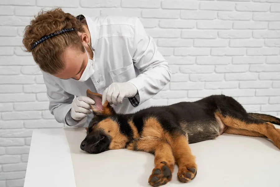
Demodex can also cause localised ear inflammation.
Complications and consequences
Generalised demodicosis often involves secondary bacterial infection. Infection can progress to deep folliculitis or furunculosis. The skin may then have crusts, discharge, ulcers and draining tracts. Severe disease can involve enlarged lymph nodes, fever and tiredness. In the most serious cases, bacterial infection can cause life-threatening sepsis.
The outlook for localised demodicosis is usually good, and spontaneous recovery is common. However, mild localised disease can spread and become generalised. Modern treatment gives young dogs with generalised disease a good outlook. In an older dog, any underlying illness also affects the prognosis. Recurrence during the first year is not uncommon.
Diagnosis at the vet
Diagnosis is usually confirmed by a deep skin scraping examined under a microscope. Several sites are sampled to represent the skin's condition as well as possible. Squeezing the skin during collection helps bring mites from deep hair follicles to the surface. Active lesions, such as bumps and pustules, are preferred for sampling.
Finding one mite across several scrapings can be incidental. Finding several strongly supports clinical demodicosis. If only one is found in a dog with signs, more samples are taken to confirm the diagnosis. Mite numbers and developmental stages are recorded for monitoring.
Hair samples can be taken around the eyes or between the toes when scraping is difficult. Hair examination is called a trichogram. A sample may need 50–100 hairs to improve the chance of finding mites. A negative hair sample is followed by skin scraping before demodicosis is ruled out. Some difficult paw cases or Shar Peis need a skin biopsy.
Cytology from a pustule or draining tract investigates bacterial infection. In severe deep infection, bacterial culture helps select a suitable antibiotic. Adult-onset disease also prompts investigation of an illness weakening immunity. Extensive tests do not always identify one. In one larger review, the cause remained unknown in 30% of adult dogs.
Scraping only the centre of an ulcerated area is unhelpful because mites are less readily found there. Hair may be lightly shortened so sample material is not lost in the surrounding coat. Mites can sometimes also be seen in discharge from a pustule or draining tract.
Dogs may swallow mites while licking their skin, and these may later appear on faecal examination. A negative faecal result does not rule out demodicosis. Faecal testing can also investigate other internal parasites relevant to general health.
Treatment
The vet prescribes medication. Mild localised demodicosis in a young dog can usually be monitored without mite treatment. It often resolves within 1–2 months. Monitoring checks whether patches disappear or spread. Generalised demodicosis needs treatment. Care involves reducing mites, controlling skin infection and treating any underlying illness.
Isoxazolines are now usually the first-choice medicine group for generalised demodicosis. Other mite treatments include medicines in the macrocyclic lactone group. Choice depends on your dog's individual circumstances and local prescribing rules. Epilepsy may affect whether an isoxazoline is suitable. Testing may be needed before medicine selection for the MDR1, or ABCB1, gene variant that causes sensitivity to certain macrocyclic lactones.
Topical antibacterial skin care may be enough for mild to moderate secondary bacterial infection. Oral antibiotics are therefore not automatically necessary for every dog. However, severe bacterial infection may require systemic antibiotic treatment. Corticosteroids may be contraindicated in demodicosis. The vet decides on changes to existing medication.
Response is monitored through repeat skin scrapings as well as skin appearance. Rechecks are usually monthly. Samples are taken from the same sites as previously. Mite numbers and immature developmental stages should decrease during monitoring. Stopping treatment is considered only after at least 2 consecutive negative scrapings. Some dogs need several months of treatment.
Adult-onset demodicosis may respond more slowly than juvenile disease. If both skin signs and mite counts remain unchanged, a treatment change is considered. Assessing the outcome can take weeks to months.
Prevention
Breeding selection is central to prevention: breeding is not recommended from a dog that has had generalised demodicosis. The same recommendation applies to the affected dog's parents. Some breed-club recommendations also exclude a dog that has produced affected puppies from different matings. Becoming symptom-free after treatment does not change the significance of inherited susceptibility.
Mites survive poorly away from dogs, and the environment is not generally considered an infection source. Ordinary Demodex canis demodicosis is not considered contagious. A mite on another dog's skin therefore does not mean the same thing as contagious sarcoptic mange, for example. Preventive mite medication does not replace investigation of an affected dog's underlying cause.
Important for dog owners
Photograph bald areas in the same lighting and record their locations. Note new patches, paw tenderness, itching and any general signs of illness. Bring a list of every medicine and details of how treatment has worked in practice to rechecks. Your dog may look better before the mites have disappeared. Hair regrowth alone is therefore not a suitable reason to miss an agreed appointment.
Do not try corticosteroids or another animal's mite medicine yourself. Inherited medicine sensitivity, for example, can affect suitability. Tell the vet about previous seizures and every change during treatment too. Continue watching the skin after treatment ends: at least 12 months of follow-up has been recommended before assessing permanent recovery. If new changes develop, previous response and sample results help guide further care.
Frequently asked questions
Can Demodex mites spread to people or the family cat?
Canine Demodex mites are not considered transmissible to humans. They are adapted to their host, and cross-infection between dogs and cats has not been demonstrated. Humans have their own Demodex species, so human demodicosis is different from a dog's skin disease.
Can a small bald patch heal without medicine?
Mild localised demodicosis in a young dog often resolves on its own. The patch's cause still needs identifying first. If more changes appear or spread into generalised disease, treatment needs reassessment. Generalised demodicosis requires veterinary treatment.
Why does demodicosis not always itch?
Mild demodicosis often causes little or no itching. Your dog may therefore seem well despite bald patches appearing. Itching can increase later when secondary bacterial or yeast infection develops.
Why is treatment monitored with skin scrapings?
Skin appearance may improve before mites disappear. Scrapings allow comparison of mite numbers and developmental stages with earlier visits. The vet decides when to stop from the skin and sample findings, so rechecks remain important even as hair grows back.
Can a recovered dog be used for breeding?
Breeding is not recommended from a dog that has had generalised demodicosis, even after the skin heals. Juvenile disease has an inherited basis probably involving several genes. Successful treatment does not remove this susceptibility affecting offspring risk.
Sources
- Diagnosis and Treatment of Bacterial and Parasitic Dermatologic Emergencies in Dogs, Today’s Veterinary Practice, accessed 2026-11-03.
- Mange in Dogs and Cats, MSD Veterinary Manual, accessed 2026-11-03.
- Demodex spp., Companion Animal Parasite Council, accessed 2026-11-03.
- Diagnosis and treatment of demodicosis in dogs and cats, World Association for Veterinary Dermatology / Veterinary Dermatology, accessed 2026-11-03.
- Immunologiset ongelmat: demodikoosi, Suomen Ranskanbulldogit ry, accessed 2026-11-03.
- Sikaripunkki (demodikoosi), Koirakissaklinikka, accessed 2026-11-03.
- Demodex canis, University of Saskatchewan, accessed 2026-11-03.
This information does not replace a visit to a vet. In an emergency, contact your nearest emergency vet: find a vet (RCVS).