
Dog diseases · Digestive system
Gallbladder mucocoele in dogs – symptoms and treatment
A gallbladder mucocoele is an accumulation of thick mucus and concentrated bile within the gallbladder. It may be an incidental finding without symptoms or cause abdominal pain, vomiting and deterioration in general condition. Bile duct obstruction or gallbladder rupture can make the situation life-threatening.
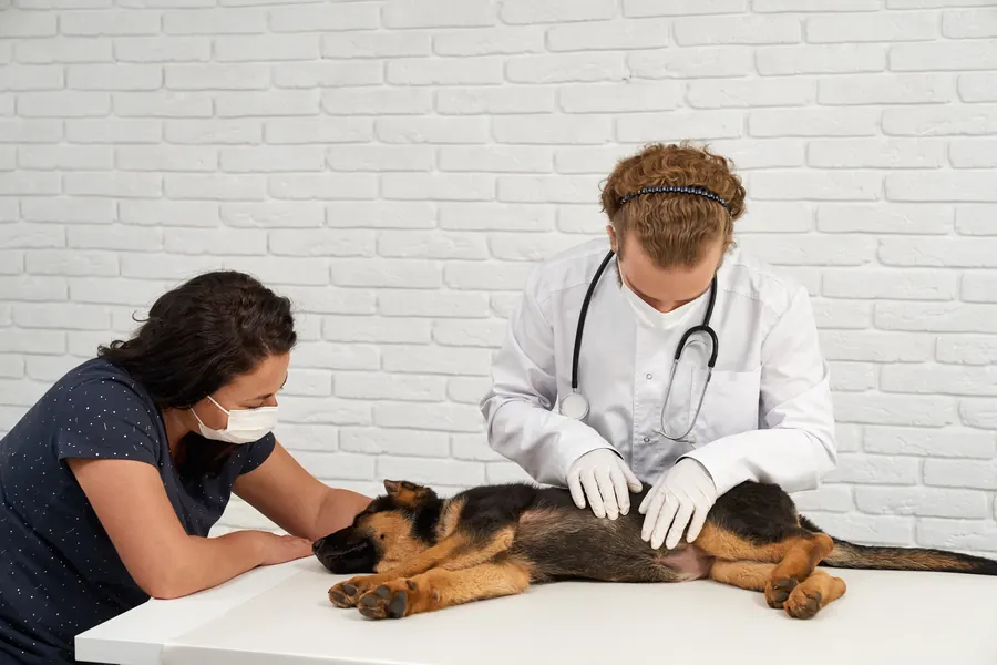
When to see a vet urgently
Seek emergency veterinary care if suspected or diagnosed gallbladder disease is accompanied by sudden deterioration. Particular warning signs include the following:
- Your dog's general condition suddenly collapses or it becomes very weak.
- Severe abdominal pain occurs with vomiting or marked weakness.
- The whites of the eyes or mucous membranes turn yellow alongside abdominal pain or fever.
- Your dog breathes rapidly, has a fever and a painful abdomen.
You can find a vet near you to arrange care.
Overview
The liver produces bile, which travels along bile ducts to the gallbladder for storage. The gallbladder concentrates bile and releases it into the small intestine around mealtimes.
In the small intestine, bile helps process dietary fats, so normal bile flow is part of digestion. With a mucocoele, gallbladder contents become thick and may adhere to the lining.
This is an important canine biliary disease that has been recognised increasingly over recent decades. In one hospital study from northern Europe involving 97 dogs with mucocoeles, 18% had no symptoms at diagnosis.
In a dog without symptoms, the change may be found during an abdominal ultrasound performed for another reason.
Causes
Bile stagnation and mucus dehydration alter the contents
Several factors contribute to mucocoele development, and not all triggers are known. Impaired gallbladder emptying leaves bile in the organ and promotes fluid absorption out of its contents.
As mucus dehydrates, its gel-forming components can become a rubbery mass that is difficult to move. The accumulating mass may extend into the bile ducts and obstruct bile flow out of the liver.
Increasing pressure can reduce blood flow to the gallbladder wall and cause tissue death. Stagnant bile and gallbladder distension predispose the wall to inflammation.
The mucus layer may also shelter bacteria if a biliary infection persists or recurs. Normal mucus hydration involves cellular CFTR channels, whose dysfunction has been linked to mucocoele formation.
This dysfunction does not mean the dog has the same CFTR gene defect as in human cystic fibrosis. Studies of an association between the ABCB4 gene and mucocoeles have produced conflicting results.
Risk factors
The disease mainly affects middle-aged and older dogs, although younger dogs can be affected too. In the northern European hospital study, the mean age at diagnosis was 9.5 years, with a range of 2.0–14.5 years.
Breed predisposition is particularly recognised in Shetland Sheepdogs, Miniature Schnauzers and Cocker Spaniels. The northern European patient group included 24 Shetland Sheepdogs, or 25% of affected dogs.
That patient proportion is not an estimate of disease risk for all Shetland Sheepdogs. No clear difference in susceptibility between sexes has been identified.
Cushing's disease, hypothyroidism and increased blood lipid levels are recognised predisposing factors. Diabetes is another disease that may occur as a predisposing underlying factor.
In a susceptible dog, a high-fat diet or glucocorticoid medication may accelerate maturation of an already developing change.
Typical symptoms
Signs may be vague and appear as intermittent abdominal upset.
- Your dog vomits; vomiting is among the most commonly observed signs.
- Appetite declines and your dog leaves meals it previously enjoyed.
- Your dog seems tired, reluctant to move or more subdued than usual.
- Abdominal pain may appear as restlessness or episodes of discomfort.
- A yellow tinge to the whites of the eyes or mucous membranes may indicate impaired bile flow.
- Some dogs have diarrhoea, fever or increased drinking and urination.
Acute illness has lasted around 5 days on average, but mild intermittent signs may occur for months. Among symptomatic dogs in the northern European group, 41% had shown signs for less than a week before diagnosis.
Signs lasting around 1–4 weeks were reported in 30% of symptomatic dogs. A further 15% had shown signs for 1–3 months before investigation.
A history exceeding 3 months was present in 14% of symptomatic dogs. Among the 80 symptomatic dogs in the northern European study, abdominal pain was reported in 71%.
In the same symptomatic group, 69% vomited, making vomiting common but non-specific.
Complications and consequences
Obstructed bile flow can cause jaundice and rapidly worsening systemic illness. Gallbladder rupture releases bile into the abdomen and can cause life-threatening bile peritonitis.
Bacterial infection and sepsis worsen the situation, particularly in dogs ill because of gallbladder rupture. A dog recovering from surgery may have a good long-term outlook.
In the northern European study, 22% of operated dogs developed a major complication and died or were euthanased. Earlier surgical patient groups reported mortality ranging from 5–40%.
Surgical mortality of 5% has been reported in dogs without symptoms. In the same comparison, surgical mortality in symptomatic dogs was 20%.
Disease stage, general condition and concurrent diseases therefore influence surgical risk. Serious surgical complications can also include pancreatitis or bleeding within the abdomen.
Diagnosis at the vet
Ultrasound distinguishes ordinary sludge from a mucocoele
Abdominal ultrasound is the primary imaging test, assessing gallbladder contents, the wall and surrounding tissues. A mature mucocoele may form a star-shaped pattern or one resembling the inside of a kiwi fruit.
Loose biliary sludge moves with gravity, whereas the jelly-like contents of a mucocoele remain in place when position changes. An irregular or discontinuous gallbladder wall and free abdominal fluid may raise suspicion of rupture.
However, rupture can be missed on ultrasound. In one clinical study, ultrasound sensitivity for detecting bile peritonitis was only 56%.
Where necessary, emptying can be measured by comparing gallbladder size before a meal and during post-meal monitoring. A healthy gallbladder's volume should decrease by at least 25% during 3 hours of monitoring after a meal.
Computed tomography has not improved assessment of gallbladder size or movement compared with real-time ultrasound.
Blood results, inflammation and possible bile leakage are investigated
Blood tests often show increased liver enzymes and sometimes an increased bilirubin level. In the northern European group, alkaline phosphatase was increased in 75% of dogs tested.
ALT was increased in 68% of dogs in which it was measured. Similar liver enzyme changes also occur in other liver and biliary diseases.
Blood cholesterol and triglycerides are checked to investigate possible lipid metabolism problems. In the northern European study, increased CRP, an inflammatory marker, did not directly correspond to bacterial growth in bile.
A liver biopsy taken during surgery may help investigate concurrent liver disease alongside the gallbladder change. Blood bilirubin within the reference range does not rule out bile leaking into the abdomen.
Abdominal fluid bilirubin at least twice the serum concentration supports a diagnosis of bile peritonitis. Passing a needle through the abdominal wall to sample the gallbladder is not a routine test for a suspected mucocoele.
A gallbladder removed at surgery is sent for histopathology to assess the change in more detail. Bile may also be collected for bacterial culture during surgery to help plan antimicrobial treatment.
In the northern European study, 85% of bile cultures were negative, so bacteria were not grown from every dog tested.
Treatment
Surgical timing depends on the findings and the dog's condition
Your vet selects treatment according to symptoms, general condition, blood tests and imaging. Gallbladder removal, or cholecystectomy, is usually the first-choice treatment for a symptomatic or complicated mucocoele.
Suspected rupture requires emergency surgery rather than waiting for a planned follow-up appointment. The gallbladder can be removed through open surgery or, in some cases, with laparoscopic assistance.
Simply emptying the contents while preserving the gallbladder is not recommended because the mucocoele usually recurs. With a ruptured gallbladder, bile and inflammatory material are cleared from the abdominal cavity.
Some of these patients also need an abdominal drain to remove fluid. An identified bacterial biliary infection is treated with an antimicrobial selected by the vet.
Severe bile peritonitis may require antimicrobial treatment after surgery.
Medical treatment requires monitoring without obstruction or leakage
Medical treatment may be considered for a stable dog if there are no signs of biliary obstruction or bile leakage. Ursodeoxycholic acid can increase bile flow and make bile acids less harmful.
However, bile duct obstruction is a contraindication to ursodeoxycholic acid. S-adenosylmethionine may be used to support liver cells at the vet's discretion.
Medication does not always prevent progression or remove the need for later surgery. Monitoring uses blood tests and repeated ultrasound examinations.
Described follow-up intervals include 6 weeks and, depending on the situation, 1–2 or 1–3 months. Worsening symptoms, blood results or ultrasound findings indicate that medical treatment is not controlling the situation adequately.
Increased blood lipids and identified hormonal diseases are treated alongside this according to their own plans. A low-fat complete diet is particularly suitable for a dog with a lipid metabolism disorder.
Some dogs still need medication after gallbladder removal. Postoperative liver values usually improve unless another ongoing disease or complication is present.
In the northern European study, postoperative hospital stays ranged from 0–4 days. The liver can be reassessed by ultrasound where needed to check whether concurrent tissue changes are resolving.
Prevention
No reliable home method prevents mucocoeles because their development involves multiple factors. Good control of identified hormonal and metabolic diseases is part of addressing risk factors.
Dietary fat is assessed in treats and chews as well as main meals. Vitamin D supplementation has not been shown to prevent mucocoeles, although low levels have been associated with the disease.
Some breed-club guidance recommends gallbladder examination at around 3–6 years of age for Shetland Sheepdogs used for breeding. Some breeding strategies exclude clinically affected dogs and those with symptoms from breeding.
The precise inheritance mechanism in Shetland Sheepdogs remains unknown despite clear breed predisposition.
Important for dog owners
Record appetite, vomiting, situations suggesting abdominal pain and changes in drinking. Bring details of the full diet, including treats, and a list of medicines and supplements. Mention previous ultrasound findings so the new examination can be compared with earlier results.
Treatment decisions are not based solely on how your dog looks at home, because a significant change may cause few symptoms.
Ask the veterinary practice for a written follow-up plan and emergency instructions. Do not alter prescribed hormonal medication or start bile-related products yourself. If your dog starts vomiting severely, becomes jaundiced or deteriorates sharply, contact an emergency vet without waiting for the next check.
Vomiting, diarrhoea or increased liver values after surgery may still need further investigation.
Frequently asked questions
Does biliary sludge always mean a mucocoele?
Loose biliary sludge usually moves with the dog's position and gravity. The firm contents of a mucocoele remain in place instead of behaving like freely moving sludge. Significance is assessed using ultrasound and the dog's other test results.
Can a dog have a mucocoele without symptoms?
Yes: in the northern European hospital study of 97 dogs, 18% had no symptoms at diagnosis. The change may be discovered during another ultrasound examination. Medical treatment still requires monitoring because the condition can progress. General condition and investigation findings are assessed together when planning care.
Must the gallbladder always be removed immediately?
A ruptured gallbladder or bile duct obstruction may require urgent surgery. In a stable situation without leakage or obstruction, medication and planned monitoring may be considered. Worsening results are a reason to reassess the need for surgery. Medication does not always stop progression.
Can a dog live without a gallbladder?
A dog can live after gallbladder removal, and the long-term outlook for one that survives surgery may be good. The liver continues producing bile even when its storage organ has been removed. Some dogs need ongoing medication or monitoring of gastrointestinal signs. Vomiting and diarrhoea, for example, can persist after surgery.
Can low-fat food replace treatment?
A low-fat complete diet may help, particularly when blood lipids are increased. However, it does not repair an already ruptured gallbladder or remove the need for urgent care caused by biliary obstruction. Diet forms part of the vet's overall plan.
Sources
- Canine Gallbladder Mucocele, Merck Veterinary Manual, accessed 2026-11-03.
- Retrospektiivinen tutkimus: koiran sappirakon mukoseele Suomessa – elinajan ennusteeseen ja kuolemanriskiin vaikuttavat tekijät, Suomen Eläinlääkärilehti, accessed 2026-11-03.
- Sappirakon mukoseele, Shetlanninlammaskoirat ry, accessed 2026-11-03.
- Tutkimushankkeet: sappirakon poisto koiralla, Helsingin yliopisto, accessed 2026-11-03.
- Mikä on mukoseele?, Pyynikin eläinlääkärit, accessed 2026-11-03.
- How an Emerging Disease in Dogs is Shedding Light on Cystic Fibrosis, NC State University, accessed 2026-11-03.
This information does not replace a visit to a vet. In an emergency, contact your nearest emergency vet: find a vet (RCVS).
More on this topic: Digestive system
- Echinococcus tapeworms in dogs – infection and echinococcosis
- GDV in dogs: recognising a twisted stomach emergency
- Giardia in dogs: symptoms, tests and treatment
- EPI in dogs: pancreatic insufficiency symptoms and treatment
- Pancreatitis in dogs: symptoms, tests and treatment
- Mould poisoning in dogs: mouldy food and compost