
Dog diseases · Skin and coat
Histiocytoma in dogs: a red lump on the skin
A histiocytoma is usually a benign tumour formed from cells of the skin's immune system. It often appears on a young dog's skin as a rapidly growing red lump. Most disappear on their own within 2–3 months. A cell sample or biopsy confirms the diagnosis when needed.
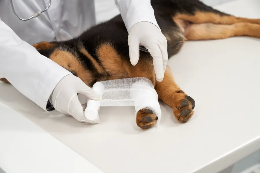
When to see a vet urgently
Contact a vet promptly if the lump causes problems or your dog's general condition changes. Feeling unwell alongside a skin change needs prompt assessment.
- The lump bleeds repeatedly or bleeding does not settle.
- Its surface becomes ulcerated, inflamed or painful.
- Your dog becomes tired or loses their appetite.
- A suture in the removal wound comes out, or the wound swells markedly.
You can find a vet near you to arrange care.
Overview
Histiocytoma is a common skin tumour in dogs. Its cells resemble the skin's Langerhans cells, which contribute to the body's defences. The growth is typically a single, raised, hairless lump. Common sites include the head, ear flaps and limbs. The lump usually moves freely relative to surrounding tissues.
Rapid growth does not automatically mean malignancy in this tumour type. Cells may multiply quickly during the first 1–4 weeks. Later, your dog's own immune system may cause the tumour to disappear. An ordinary skin histiocytoma should not be confused with other diseases arising from histiocytes. Histiocytic sarcoma, for example, is a different tumour that can follow a very different course.
In the Lisbon and Setúbal regions of Portugal, about 4.0 histiocytoma cases per ten thousand dogs were recorded annually. This figure is based on cases confirmed by tissue samples, so lumps resolving on their own without sampling may be missed. It should not be treated as a universal estimate of risk.
Causes
Langerhans cells are part of the skin's immune surveillance. They take up foreign antigens, structures recognised by the immune response. Such structures come from sources including pollen and microorganisms. However, this normal function does not establish what triggered a particular dog's lump to grow.
The cells can move to local lymph nodes and present the structures they have recognised to other immune cells. T lymphocytes activated in this way help protect the body. Spontaneous regression of a histiocytoma is associated with an immune reaction against the tumour. It is not a tumour transmitted between dogs.
T cells that destroy tumour cells may also become activated as a histiocytoma regresses.
Abnormal immune regulation is suspected to contribute to histiocytoma development. Genetics may also play a part because some breeds have a higher risk. However, no single factor definitely causing the tumour is known.
Risk factors
Histiocytomas are typically seen in dogs younger than 3.5 years, but can occur in older dogs too. Of 359 tissue-confirmed Portuguese cases, 48.8% involved dogs aged 2 years or younger. Adult age therefore does not rule out the diagnosis. Histiocytomas can occur in any breed.
Susceptible breeds include Boxers, English Bulldogs and Boston Terriers. Dachshunds and Great Danes are also described as more susceptible than usual. In the Portuguese population comparison, French Bulldogs had about 11.5 times the risk of crossbreeds. The corresponding risk ratio for Boxers was about 8.3.
In the 359 confirmed Portuguese cases, ages ranged from 2 months to 13 years. Males accounted for 58.5% of cases. Females accounted for 41.5%. These were tissue samples submitted to one local laboratory, so the distribution should not be directly applied to dog populations elsewhere.
Typical symptoms
A lump can appear quickly and grow visibly over a short time. Typical size is about 0.2–3.0 cm. Histiocytomas can occur anywhere on the skin, although the head and limbs are common sites. Features you may notice at home include:
- A rounded, button-like raised growth.
- A surface that is usually red.
- Hair often missing over the lump.
- A smooth surface.
- Ulceration or crusting of the lump.
- Licking, biting or scratching of an irritated area.
- Bleeding or infection in an ulcerated lump.
However, not every raised skin lump has the same diagnosis. Mast cell tumours and plasma cell tumours, for example, can resemble histiocytomas.
A histiocytoma is often clearly defined against surrounding skin. The average lump diameter in the Portuguese cases was about 1.6 cm. By comparison, a viral papilloma may have a hardened, cauliflower-like surface. A histiocytoma's usual smooth surface differs, but appearance does not replace sampling when needed.
A histiocytoma is often noticed while stroking or grooming a dog. A typical lump may cause no discomfort. However, swelling can develop around an inflamed lump. An ulcerated surface may discharge pus if infection develops.
Complications and consequences
A typical histiocytoma is benign, but a damaged surface can develop secondary infection. Itching and licking can worsen local inflammation. If a lump is repeatedly rubbed or injured, it may need removal. Removal provides a permanent cure in most cases.
A local lymph node may sometimes enlarge because of secondary infection. Enlargement can also indicate a different diagnosis or spread of abnormal cells. Several tumours appearing together, or later at different sites, are unusual with ordinary histiocytoma. Such a situation needs reassessment.
Diagnosis at the vet
The vet examines the lump's size, shape, location and texture. A button-like appearance may suggest histiocytoma. However, a precise diagnosis relies on microscopic examination of cells or tissue. Histiocytoma can often be recognised through cytology, meaning a cell sample.
Fine-needle sampling collects cells from the lump with a small needle and spreads them onto a slide. The sample can often be taken without general anaesthesia. Your dog is sedated if needed for safe collection. Cytology quickly provides preliminary information about the growth.
Histiocytomas are round-cell tumours, which often yield cells readily on fine-needle sampling. The microscope typically shows round or oval cells of fairly similar size. Lymphocytes, a type of immune cell, may also be present. Distinguishing different round-cell tumours affects assessment of the outlook and further investigations.
If cytology does not give an adequate answer, a biopsy is taken. A small growth may be removed completely as the sample. In histopathology, a pathologist examines tissue structure. A removed tumour is also assessed for complete excision.
A histiocytoma can be difficult to distinguish from other tumours even by tissue appearance. Conditions to distinguish include inflammatory granuloma, mast cell tumour, plasma cell tumour and skin lymphoma. Sedation is used for biopsy collection when necessary. The skin sampling site may be closed with a small suture.
A cell sample spread on a slide is stained so cell structures can be identified under the microscope.
Treatment
The vet prescribes treatment and any medicines. A confirmed histiocytoma causing no problems can be monitored while awaiting regression. Most tumours regress within about 2–3 months without treatment. However, ulceration, itching, infection or bleeding may need treatment during monitoring.
Antibiotics may be needed for secondary bacterial infection. They are not routine treatment for every histiocytoma. If the lump has not begun shrinking within 1–2 months, the vet may recommend removal. Removal is also considered when ulceration or bleeding cannot otherwise be controlled.
A surgically removed lump can be sent to a pathologist to confirm histiocytoma. The examination also checks surrounding surgical margins for tumour cells. If it is another tumour type, further treatment is planned according to the final diagnosis. Tumour type and location influence treatment choice.
Keep a surgical wound clean and dry. Prevent licking, scratching and biting while it heals. An ulcerated lump that has not been removed is also kept clean. Sutures coming out or marked swelling need veterinary assessment.
During skin wound healing, exercise is usually restricted for 1–2 weeks or until suture removal. Running and jumping can put too much strain on the wound. Swimming and bathing are also avoided until the wound closes. Moisture can carry bacteria into the wound and increase infection risk.
Removable skin sutures are usually taken out 7–14 days after surgery. Timing depends on the procedure performed. Sometimes a wound is closed with dissolvable sutures beneath the skin that do not need removal. Pain relief may be prescribed for home use after surgery.
Prevention
The cause of histiocytoma formation is incompletely understood. However, preventing scratching and licking of an existing lump reduces ulceration, bleeding and infection. Showing a new skin change to a vet is part of early problem recognition.
There is no need to avoid other dogs because of infection fears with a histiocytoma: spread between animals has not been reported. Regular skin checks help you notice new lumps. Avoid breaking the skin when handling an existing growth. An ulcerated area needs to be kept clean even when the tumour itself has been confirmed benign.
A histiocytoma on a limb can rub against the ground and become irritated. Soft bedding may protect it from this friction. There is no home treatment that removes the tumour: normal spontaneous regression relies on the dog's own immune response.
Important for dog owners
Photograph the lump and note the date it was first seen. Use the same distance and place a small measuring scale beside it in comparison photographs. Record when growth stops or the lump starts shrinking. Tell the vet if your dog rubs the area, the lump bleeds or other skin changes appear.
Ask whether the diagnosis has been confirmed by cytology or biopsy. If monitoring is chosen, agree in advance which changes mean another visit is needed. Keep the pathology report for future reference. Do not apply covering or irritating products yourself, as the growth's surface needs to remain assessable.
A collar or protective suit may prevent your dog from biting the surgical site. Wound edges should remain together. A bad smell or discharge from the wound is a reason to contact a vet.
Frequently asked questions
Is a histiocytoma cancer?
An ordinary canine skin histiocytoma is a benign tumour. However, a similar lump may be a different skin tumour, so diagnosis may require cytology or a biopsy. Red colour or a button-like appearance alone is not enough reason to leave a new lump unexamined.
How quickly does a histiocytoma disappear?
Most histiocytomas disappear on their own within 2–3 months without treatment. If a lump does not start shrinking within 1–2 months or develops troublesome ulceration, the vet may recommend removal. Recurring pain or bleeding is assessed without waiting until the monitoring period ends.
Can a lump in an old dog be a histiocytoma?
Yes. Histiocytomas occur in dogs of all ages, although they are more common in young dogs. Age alone therefore neither confirms nor excludes the diagnosis. A new lump in an older dog also needs examination and sampling to establish its type when necessary.
Can histiocytoma spread to other dogs?
Spread of histiocytoma between animals has not been reported. It is therefore not managed for infection risk in the same way as viral warts. Scratching or licking can still damage the surface and cause local inflammation.
Why is tissue examination of a removed lump worthwhile?
Tissue examination confirms the type of lump and helps assess the outlook. It can also show whether tumour extends to the excision margin. The result is particularly useful when a preliminary assessment turns out to be something other than an ordinary histiocytoma.
Sources
- Tumors of the Skin in Dogs: Histiocytic Cell Tumors, MSD Veterinary Manual, accessed 2026-11-03.
- Skin lumps and tumours in dogs, PDSA, accessed 2026-11-03.
- Koiran ihomuutosten diagnosointi ja hoito, Eläinlääkärikeskus SoVet, accessed 2026-11-03.
- Lymphocytic, Histiocytic, and Related Cutaneous Tumors in Animals, MSD Veterinary Manual, accessed 2026-11-03.
- Cutaneous Histiocytoma in Dogs, VCA Animal Hospitals, accessed 2026-11-03.
- Color Atlas of Canine Cutaneous Round Cell Tumors, MSPCA-Angell, accessed 2026-11-03.
- Epidemiology of Canine Cutaneous Histiocytoma: Insights From Age and Topographic Clustering, Veterinary Dermatology, accessed 2026-11-03.
- Post-Operative Instructions in Dogs, VCA Animal Hospitals, accessed 2026-11-03.
- Histiocytoma in Dogs: Symptoms and Treatment, PetMD, accessed 2026-11-03.
This information does not replace a visit to a vet. In an emergency, contact your nearest emergency vet: find a vet (RCVS).
More on this topic: Skin and coat
- Acne in dogs: chin spots and treatment
- Alopecia X and seasonal flank alopecia in dogs
- Paw pad injuries in dogs: first aid and wound care
- Atopic dermatitis in dogs: itching, tests and long-term care
- Demodectic mange in dogs: Demodex mites and skin disease
- Fibrosarcoma in dogs: symptoms, diagnosis and treatment