
Dog diseases · Eyes, ears and mouth
Keratitis in dogs: corneal inflammation and treatment
Keratitis means inflammation of the clear surface at the front of the eye. Ulcerative inflammation can cause sudden pain and excessive watering. In a slowly progressing form, you may first notice discolouration spreading from the edge of the cornea.
When to see a vet urgently
Have a painful eye or one with a newly cloudy surface examined promptly. Contact an emergency vet immediately if your dog's eye develops any of the following sudden changes.
- Your dog cannot open the eye because of pain or squints constantly.
- The cornea rapidly becomes cloudy, or a distinct pale patch appears on its surface and the eye is painful.
- The eye has been injured, for example by a sharp object.
- Your dog suddenly cannot see normally.
- The eye's surface looks indented or its shape changes noticeably.
Overview
What happens in the cornea?
The cornea lets light into the eye and protects its internal structures from the environment. Its surface is covered by the epithelium, which is several cell layers thick. Most of the cornea's thickness consists of the stroma beneath this surface layer. A thin layer called Descemet's membrane lies between the stroma and the innermost cell layer.
A superficial ulcer affects the corneal epithelium. A deep ulcer can extend through the stroma as far as Descemet's membrane. The cornea can also become inflamed and develop blood vessels without an ulcer on its surface. Cloudiness may result from swelling, inflammatory cells, pigment or the formation of fibrous tissue.
Causes
Friction, dryness and infection
Keratitis affecting only one eye is often caused by an injury. A foreign body can damage the cornea and sustain inflammation until it is removed. The edge of an inward-turning eyelid can rub the eye's surface. Extra eyelashes growing from the eyelid margin or inner surface can also scratch the cornea. If the eyelids do not close properly, tears cannot spread over the whole surface.
In dry eye, the watery component of tears may be insufficient, predisposing the eye's surface to inflammation. Bacteria normally living on the eye's surface can enter a damaged area when the protective epithelium breaks down. When 476 dogs in the United States were sampled for suspected bacterial keratitis, staphylococci accounted for 32.3% of the bacteria isolated. Streptococci accounted for 19.1% of bacterial findings. Pseudomonas species accounted for 12.5%.
The yeast Candida albicans was identified as the cause of inflammation in both eyes of a 4-year-old Chihuahua.
Immune-mediated inflammation and pigment
In pannus, immune cells enter the cornea from its edge. Pannus is chronic superficial keratitis that usually progresses in both eyes. Punctate keratitis is an inherited immune-mediated disease causing small, pinpoint corneal ulcers. In pigmentary keratitis, prolonged irritation can lead to pigment accumulating in the corneal surface and front of the stroma. Pigmentary keratitis can also occur without an obvious anatomical explanation.
Risk factors
Protruding eyes in short-muzzled dogs can predispose them to corneal injury. A nasal skin fold can predispose a dog to corneal ulceration, particularly in short-muzzled breeds. A Pug's cornea may have reduced sensitivity. In a study of dogs attending primary veterinary practices in England in 2013, corneal ulcers were recorded in 834 dogs (0.80%). In that study, short-muzzled dogs had an odds ratio of 11.18 for corneal ulceration compared with crossbreeds. Corneal ulcers were recorded in 5.42% of Pugs. The proportion in Boxers was 4.98%. In Shih Tzus, it was 3.45%.
Breeds predisposed to pannus include German Shepherd Dogs, Belgian Shepherd Dogs of the Tervueren variety and Border Collies. Punctate keratitis occurs particularly in Long-haired Dachshunds, in which changes may be detected at 1.5–4 years of age. Among 210 Pugs examined at events in Britain, 193 had pigmentary keratitis in at least one eye (91.9%). Of those 193 affected Pugs, 176 had changes in both eyes (91.2%). Pigmentary changes were more common in older Pugs. They were more severe in Pugs with greater inward rolling of the inner corner of the lower eyelid. One northern European Pug breeding report recorded keratitis in 9 of 47 examined Pugs.
Typical symptoms
Pain from a corneal ulcer is often more apparent than the ulcer itself.
- Your dog squints or keeps the eye closed.
- The eye produces more clear tears than usual.
- Your dog avoids bright light.
- Swelling may make the cornea look bluish and cloudy.
- The eye may produce discharge containing mucus and pus.
- Fine, branching blood vessels may grow onto the eye's surface.
Pannus often begins with redness and superficial blood vessel growth at the corneal edge. Changes caused by pannus may differ in severity between the eyes. Pannus may be accompanied by thickening and loss of colour along the third eyelid margin. Pigmentary keratitis often appears as a dark area spreading from the inner corner towards the centre of the cornea.
Complications and consequences
Melting ulcers and impaired sight
In a melting corneal ulcer, enzymes from bacteria and inflamed tissue break down the stroma. A deep ulcer can perforate the cornea. The iris may protrude into the defect through a perforated ulcer. A deep corneal ulcer can also cause inflammation inside the front of the eye.
A British hospital investigated melting corneal ulcers in 106 dogs during 2014–2018. Eye removal was necessary in 14 cases (13%) when tissue breakdown continued despite treatment. The likelihood of losing the eye was higher when only Pseudomonas grew from the ulcer. Eight of the removed eyes had a pure culture of Pseudomonas aeruginosa.
In untreated pannus, blood vessels, pigment and scarring may cover the cornea so that light cannot pass through. Pigmentary keratitis can also impair sight if the dark area spreads in front of the pupil.
Diagnosis at the vet
Examining and staining the cornea
Your vet examines the cornea closely with a magnifying slit lamp and assesses the depth of any ulcer. They also check whether an eyelid or foreign body is rubbing the cornea. Fluorescein dye adheres to areas where corneal epithelium is missing. Under cobalt-blue light, the stained area appears green. Fluorescein can also help check whether fluid is leaking from inside the eye through the cornea. The Schirmer tear test measures production of the watery tear component over 1 minute.
Pannus changes typically begin at the corneal edge and may progress towards the centre, so their location helps identification. A pigmented surface may also be associated with dry eye, which can cause inflammation and thick discharge.
When is a sample needed?
A dense pale deposit at the edge or base of an ulcer may indicate bacterial infection of the damaged area. A deep or melting corneal ulcer may be sampled for bacterial culture and susceptibility testing. Cytological examination of an ulcer sample can help guide antimicrobial selection. Among 476 dogs with suspected bacterial keratitis, 220 cultures showed no growth (46.2%).
Of 112 corneal ulcers examined in Berlin during 2021–2024, 89 yielded microbial growth on culture (79.5%). Sixteen of the samples without growth came from eyes already treated with antibiotic drops (69.6%).
Treatment
Treating ulcerative keratitis
Your vet addresses the cause of ongoing corneal irritation, such as an eyelid abnormality or a foreign body lodged on the surface. A topical antimicrobial may be used for a superficial ulcer to prevent bacterial infection. For a deep or melting ulcer, the antibiotic choice is guided by the susceptibility of bacteria identified on culture. Corticosteroid drops must not be used in an ulcerated eye. Your vet may use pain relief to treat pain caused by corneal damage.
When at least 50% of the stromal thickness has been lost, a vet with expertise in ophthalmology assesses the need for surgery. One way to support a weakened cornea is to cover the defect with a conjunctival graft. In the British study of melting ulcers, 63 eyes received a graft alongside medical treatment (57%). Graft healing is monitored with repeated eye examinations. Scarring and pigment may remain in the cornea after grafting.
An ulcer in the centre of the cornea develops blood vessels more slowly than one near the edge. A superficial ulcer may heal in 3–5 days if there is no irritation or infection delaying healing. Healing of a superficial ulcer may also take 7–14 days. If the epithelial edge remains loose and the ulcer does not close, it may be a spontaneous chronic corneal epithelial defect, or SCCED. In this condition, your vet may remove loose surface tissue so that new epithelium can attach.
After removal of corneal surface tissue, a soft contact lens may be used to protect the healing surface.
Pannus, dry eye and pigmentary keratitis
Topical anti-inflammatory treatment for pannus usually continues throughout the dog's life. Your vet may prescribe ciclosporin or a topical corticosteroid for pannus once ulceration has been ruled out. Treatment can reduce corneal inflammation and blood vessel growth even if pigment remains on the surface. In one treatment follow-up, conjunctival redness associated with pannus decreased within 1 week. In the same follow-up, corneal pigment disappeared completely in 3 dogs by 6 months.
If pannus worsens when treatment is interrupted, the inflammation may be harder to bring under control again.
Artificial tears can protect the eye's surface in dogs with dry eye. An eye medicine that increases tear production may also reduce inflammation associated with dry eye. In pigmentary keratitis, an eyelid abnormality at the inner corner can be corrected if it sustains irritation.
Prevention
When an eyelid margin constantly rubs the cornea, correcting the cause reduces ongoing surface damage. Treating dry eye protects the surface from drying and associated ulcers. Ultraviolet radiation can worsen pannus. If needed, a dog with pannus can wear sunglasses designed for dogs outdoors. Greater UV exposure in dogs living at high altitude can make pannus harder to control. Breeding from a dog diagnosed with inherited pannus is not recommended. Dogs with punctate keratitis should also be excluded from breeding.
Important for dog owners
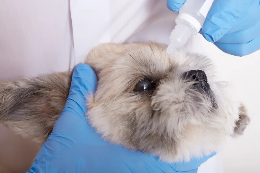
If eye symptoms recur, do not restart old corticosteroid drops on your own. A protective collar can prevent pawing and rubbing of a painful eye while it heals. When giving prescribed eye drops, keep the bottle tip away from the eye's surface and eyelids. Another person can hold your dog calmly in place while you give the drop. Your vet assesses healing of the corneal damage at a follow-up examination.
Reward your dog after giving eye drops so that the experience feels more familiar next time. Contact your practice if pain or cloudiness worsens before the scheduled check.
Frequently asked questions
Is keratitis the same as conjunctivitis?
In keratitis, inflammation affects the clear front surface of the eye, the cornea. The conjunctiva covering the white of the eye may also look red when the actual damage is in the cornea. Staining helps identify damage involving the corneal epithelium.
Could a dark patch on the eye be keratitis?
In pigmentary keratitis, dark pigment accumulates in the superficial corneal layers. The pigmented area may grow from the inner corner towards the centre of the cornea. Sight may deteriorate if pigment spreads directly in front of the pupil and blocks part of the light's path.
Why recheck the eye if my dog has stopped squinting?
At the follow-up visit, your vet checks whether the corneal damage is healing as expected. Fluorescein staining reveals whether an open area remains in the epithelium. If a corneal ulcer is deep or fails to heal as expected, your dog may be referred to a vet with expertise in eye disease.
Can keratitis be painless?
The corneal changes of pannus are usually painless because no ulcer forms on the surface. Nevertheless, inflammation can progress from the corneal edge towards the centre. Without treatment, changes can impair sight as pigment, scarring and blood vessels cover the cornea.
Can I use eye drops prescribed last time?
Drops containing a corticosteroid must not be used in an ulcerated eye. New squinting or cloudiness of the corneal surface requires a prompt eye examination. At the appointment, fluorescein staining helps establish whether the corneal surface is damaged on this occasion.
Sources
- The Cornea in Animals (updated July 2026), Merck Veterinary Manual, accessed 2026-11-07.
- Silmäsairaudet, Helsingin yliopisto, accessed 2026-11-07.
- Silmäsairaudet ja -tutkimukset, Suomen Kennelliitto, accessed 2026-11-07.
- Koiran ja kissan sarveiskalvon tulehdus (ulseratiivinen), Ruokavirasto, accessed 2026-11-07.
- Koirien sarveiskalvohaavaumat, Koirakissaklinikka, accessed 2026-11-07.
- Deep Stromal Corneal Ulcers, Descemetocele, and Iris Prolapse in Small Animals, Merck Veterinary Manual, accessed 2026-11-07.
- Pigmentary keratitis in pugs in the United Kingdom: prevalence and associated features, BMC Veterinary Research, accessed 2026-11-07.
- Mopsien jalostuksen tavoiteohjelma 2023–2027, Suomen Mopsikerho, accessed 2026-11-07.
- Patterns of Bacterial Infection and Antibiotic Resistance in Canine Corneal Ulcers: A Retrospective Analysis (Berlin, Germany, 2021–2024), Veterinary Medicine and Science, accessed 2026-11-07.
- Prevalence and Antibiotic Susceptibility of Bacterial Isolates From Dogs With Ulcerative Keratitis in Midwestern United States, Frontiers in Veterinary Science, accessed 2026-11-07.
- Melting corneal ulcers (keratomalacia) in dogs: A 5‐year clinical and microbiological study (2014–2018), Veterinary Ophthalmology / Crossref, accessed 2026-11-07.
- A case of Candida albicans-induced fungal keratitis in a Chihuahua dog, Journal of Veterinary Science, accessed 2026-11-07.
- Keratoconjunctivitis sicca (KCS) in dogs, Cornell University College of Veterinary Medicine, accessed 2026-11-07.
- Corneal ulcerative disease in dogs under primary veterinary care in England: epidemiology and clinical management (2017), Canine Genetics and Epidemiology / PubMed Central, accessed 2026-11-07.
- Evaluation of corneal ulcer type, skull conformation, and other risk factors in dogs: A retrospective study of 347 cases, Canadian Journal of Veterinary Research, accessed 2026-11-07.
- Clinical assessment of chronic superficial keratitis in dogs (2012–2019), full article, VETERINARIA, accessed 2026-11-07.
- Pannus, American College of Veterinary Ophthalmologists, accessed 2026-11-07.
- Applying Eye Drops to Dogs, VCA Animal Hospitals, accessed 2026-11-07.
- Corneal Ulcers, American College of Veterinary Ophthalmologists, accessed 2026-11-07.
- ECVO Manual: Dachshund, European College of Veterinary Ophthalmologists, accessed 2026-11-07.
This information does not replace a visit to a vet. In an emergency, contact your nearest emergency vet: find a vet (RCVS).