
Dog diseases · Skin and coat
Melanoma in dogs: symptoms, diagnosis and treatment
Melanoma is a malignant tumour originating in pigment-producing cells called melanocytes. In dogs, it can develop in sites including the mouth, skin, toes or eyes. Location strongly influences how the tumour grows and spreads.
When to see a vet urgently
Arrange a veterinary appointment if you notice a new oral lump or a painful toe lesion that does not heal.
- Contact an emergency vet immediately if your dog's mouth bleeds heavily.
- Seek emergency care if breathing becomes difficult.
- Severe pain requires an emergency assessment.
- An eye causing severe squinting needs emergency examination.
- A rapidly deteriorating general condition requires immediate assessment.
Overview
Melanocytes produce melanin, which gives tissues a dark colour. A melanocytoma is a benign tumour arising from the same cell type as melanoma. In haired skin, benign melanocytic tumours are diagnosed more often than melanomas. The oral lining and nail bed beneath a claw are common sites of malignant melanoma.
Melanoma is the most common malignant oral tumour in dogs. Oral melanoma has been estimated to account for 14.4–45.5% of all canine oral tumours. Cutaneous melanoma accounts for 0.8–2% of canine skin tumours. Melanocytic tumours of the iris are often benign.
Causes
Changes in a pigment cell
The tumour develops when melanocytes begin multiplying abnormally. Tumour cells may contain abundant dark melanin, but some melanomas have no visible pigment. Canine melanoma has not shown the same association with UV radiation as common human skin melanomas. Changes in proteins involved in regulating cell growth have been found in melanoma tissue. Some melanomas also have changes in the NRAS and KRAS genes, which relay growth signals within cells. The direct relationship between altered growth-regulating proteins and malignant transformation remains unknown. Gains and losses of chromosome segments have also been observed in malignant melanocytic tumours.
In one study of 95 tumours, no BRAF alteration was identified in any oral or cutaneous melanoma.
Risk factors
Melanoma is most commonly diagnosed in middle-aged and older dogs. In a Japanese hospital study of 123 dogs, median age at diagnosis was 12.4 years. Of these cases, 113 originated in the mouth.
Oral melanoma occurs more often in breeds including Scottish Terriers, Golden Retrievers and Dachshunds. Breeds with increased nail bed melanoma risk include Schnauzers, Rottweilers and Irish Setters. Melanocytic tumours are diagnosed more often in heavily pigmented breeds. Iris melanocytic tumours can also occur in young dogs. Dogs examined at one guide dog centre were 0.5–3.1 years old.
Typical symptoms
Oral melanoma can grow on the gum, lip, tongue or hard palate. A small oral tumour may be found incidentally during dental treatment even if your dog eats normally.
- Bad breath can be a sign of oral melanoma.
- Your dog may drool more than usual.
- Bleeding may occur from the mouth.
- An oral tumour can make eating or swallowing difficult.
- A painful, non-healing lesion may develop beside a claw.
- A nail bed tumour can cause limping or toe licking.
Cutaneous melanoma may appear as a rapidly growing nodule with an ulcerated surface. A melanocytic tumour inside the eye may appear as a dark, raised iris lesion.
Complications and consequences
Oral melanoma can invade surrounding jawbone. Oral and nail bed melanomas metastasise particularly to regional lymph nodes and lungs. Spread to the lungs can cause breathing difficulties.
A tumour growing inside the eye can inflame the iris. It can also cause glaucoma, in which eye pressure rises. Blood may accumulate in the front of the eye. A nail bed tumour can damage the toe bone. After surgery, a tumour may recur at its original site. Following radiotherapy for oral melanoma, late adverse effects were recorded in 17 of 76 dogs followed for at least 3 months. Bone necrosis is a possible adverse effect of jaw radiotherapy. Oral surgical wound problems include suture breakdown and abnormal drooling.
Diagnosis at the vet
Sampling the tumour
Your vet assesses the tumour's location, size and attachment to surrounding tissue. Dark colour alone is insufficient to identify melanoma. A fine-needle sample may reveal melanin-containing tumour cells. A poorly pigmented tumour usually requires a biopsy for identification.
If an oral tumour's surface is ulcerated or necrotic, a biopsy is taken sufficiently deep into living tissue. Immunohistochemical staining lets a pathologist identify melanocytic markers even in a pale tumour. Ki-67 staining provides information about how actively tumour cells divide. The pathologist also assesses the number of dividing cells, abnormal cell appearance and invasion into surrounding tissue. The thickness of a cutaneous melanocytic tumour can also be measured in a tissue sample. Nail bed tumour signs can resemble nail bed inflammation or digital squamous cell carcinoma.
Assessing spread
Oral melanoma TNM staging uses tumour size, lymph node findings and metastases. In stage 1, the longest tumour diameter is under 2 cm and no metastases are identified. Stage 2 includes a 2–4 cm tumour without identified spread. Stage 3 includes a tumour over 4 cm or regional lymph node spread without distant metastases. In stage 4, distant metastases are present, for example in the lungs.
A normal-sized lymph node can contain metastases. In a study of 100 dogs with oral melanoma, melanoma cells were found in mandibular lymph nodes in 53%. A dog may have 2–5 mandibular lymph nodes in the same region, and any of them can contain metastases. A lymph node can be sampled for cytology or examined as a tissue specimen if needed. Nodes can also contain melanin-laden immune cells that must be distinguished from tumour cells.
Chest X-rays or CT look for possible lung metastases. CT detects small lung nodules more sensitively than X-rays. Jaw CT assesses bone invasion and helps plan surgery.
Treatment
Surgery and radiotherapy
Your vet plans treatment using tumour location, tissue findings and extent of spread. Surgery aims to remove the tumour with enough surrounding healthy tissue. Removing melanoma invading the jawbone may require removal of part of the jaw. A pathologist examines the surgical margins to determine whether tumour reaches the edge of the removed tissue. Removal of the affected toe is usually recommended for nail bed melanoma.
Radiotherapy can be used if complete removal is impossible. When surgery is not feasible, radiotherapy can also relieve symptoms from a large tumour. A visible oral tumour may shrink within 2–3 weeks of radiotherapy. In a study of 100 dogs receiving radiotherapy, median overall survival was 294 days.
Medication and immunotherapy
Systemic treatment may be considered after surgery if metastatic risk is high. Adding chemotherapy to surgery or radiotherapy has not been shown to significantly extend survival in oral, cutaneous or digital melanoma.
A therapeutic vaccine developed for oral melanoma aims to stimulate an immune response against melanoma cells. In one study, over half the vaccinated dogs lived longer than 437 days, compared with a median survival of 324 days in a historical comparison group. In another retrospective study of 45 dogs, vaccination was not associated with longer survival.
A newer study of 150 dogs assessed antibody treatment targeting the immune system's PD-1 checkpoint. At the first response assessment, tumours shrank or disappeared in 14.7% of dogs. Treatment-associated adverse effects occurred in 40.0% of study dogs.
Melanocytic eye tumours
In some cases, a small pigmented iris lesion can be monitored with eye examinations. A well-defined iris tumour can sometimes be removed with part of the iris. In 13 dogs operated on at a guide dog centre, the tumours were benign melanocytomas. Sight was retained in the operated eyes during follow-up of up to 6.2 years. Advanced intraocular melanoma may require eye removal. In one study of 244 intraocular melanocytic tumours, 188 were benign melanocytomas and 56 were malignant melanomas.
Recovery and outlook
Most dogs can eat early after jaw surgery. After surgery, follow-up assesses local tumour control and possible metastases. Visit intervals depend on your dog's condition, tumour type and treatment received.
Median survival in surgically treated stage 1 oral melanoma has ranged from 511 to 874 days. For stage 2, corresponding medians have ranged from 160 to 818 days. For stage 3, median survival after surgery has ranged from 168 to 207 days.
In a study of 70 dogs undergoing oral melanoma surgery, median survival was 723 days. Thirty-six dogs in that surgical group had stage 1 disease. In another surgical study of 151 dogs, median survival was 346 days.
Among 87 dogs operated on for malignant melanoma of haired skin, median survival was 1,363 days. In that study, 21.8% developed metastases after surgery. For nail bed or digital melanoma without identified metastases, median survival after toe removal has been about 12 months.
Prevention
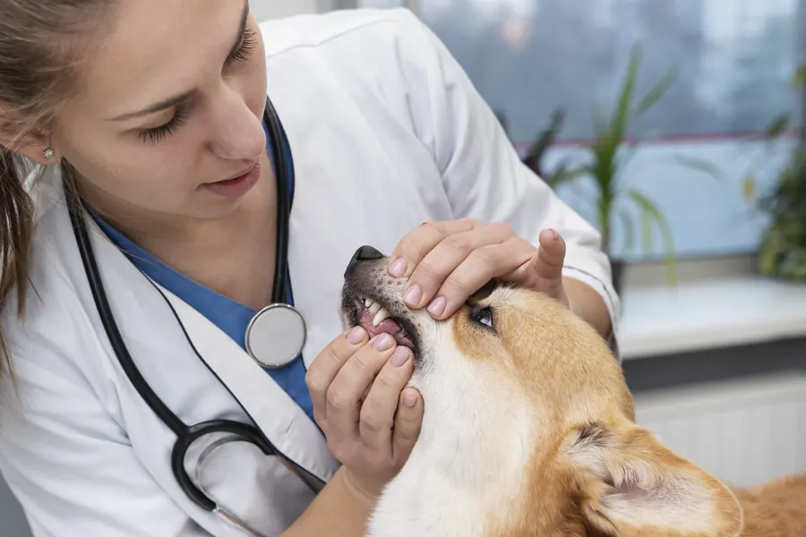
Canine melanoma has no established UV association that could provide a target for prevention. Annual veterinary health checks and regular dental care support early detection. Examining the mouth helps identify gum, lip and tongue lesions before eating becomes difficult. The back of the dental arches and tongue are also examined because melanoma can develop at different oral sites. Checking the coat and claws helps reveal nodules that are invisible from a distance.
Important for dog owners
Show your vet a new or growing lump even if it is pale or your dog otherwise seems healthy. Ask your vet to establish whether the tumour has invaded bone or spread to lymph nodes.
Prevent your dog rubbing a surgical wound against furniture or carpet for the first 2–3 weeks. Follow the operating vet's feeding and wound-care instructions. Attend scheduled follow-ups, which look for evidence of recurrence or metastases. Treatment selection considers your dog's quality of life and how far the tumour has spread.
Frequently asked questions
Is a dark lump always melanoma?
A dark lump may also be a benign melanocytoma, which can contain dark pigment too. Conversely, some oral melanomas are pale. Your vet can take a sample so the lump's cells and tissue structure can be examined.
How does nail bed melanoma differ from nail infection?
Both can cause pain, swelling and a non-healing lesion beside a claw. For persistent toe problems, your vet may X-ray the bone. A sufficiently deep tissue sample can establish whether a melanocytic tumour or another lesion is present.
Can a dog eat after part of the jaw is removed?
Most dogs learn to eat well after removal of part of the jaw. Surgery can permanently change the appearance of the muzzle or jaw. After extensive tongue or palate surgery, a temporary feeding tube may be needed.
Does a dark iris patch mean a malignant tumour?
A pigmented iris lesion can be a flat pigmented naevus or a raised tumour. Many iris melanocytic tumours behave benignly. An eye examination can establish whether a lesion is well-defined and confined to the iris or extends into other structures.
Why sample a lymph node that is not enlarged?
Melanoma cells can be present in a normal-sized node. Sampling investigates local spread because palpation does not always reveal metastases. Lymph node findings affect oral melanoma staging and help your vet plan suitable treatment.
Sources
- Mitä on päivystyshoito, Helsingin yliopisto, accessed 2026-11-07.
- Suun kasvaimet koirilla ja kissoilla, Eläinklinikka Tsemppi, accessed 2026-11-07.
- Koiran ihomuutosten diagnosointi ja hoito, SoVet, accessed 2026-11-07.
- Diagnosis and histopathologic prognostication of canine melanocytic neoplasms, Veterinary and Comparative Oncology, accessed 2026-11-07.
- Medical Oncology: Canine Oral Melanoma, NC State College of Veterinary Medicine, accessed 2026-11-07.
- Survival of dogs with melanoma from a referral veterinary hospital in Japan, Journal of Veterinary Medical Science, accessed 2026-11-07.
- Clinical descriptive and long-term outcome of melanocytic uveal lesions in young dogs, Veterinary Ophthalmology, accessed 2026-11-07.
- Tumors of Melanocytic Origin in Animals, Merck Veterinary Manual, accessed 2026-11-07.
- Digital Lesions in Dogs: A Statistical Breed Analysis of 2912 Cases, Veterinary Sciences, accessed 2026-11-07.
- Melanoma of the dog and cat: consensus and guidelines, Frontiers in Veterinary Science, accessed 2026-11-07.
- Ocular Neoplasia in Dogs, Merck Veterinary Manual, accessed 2026-11-07.
- Evaluating the Impact of Elective Nodal Irradiation for Dogs With Oral Malignant Melanoma Undergoing Hypofractionated Radiotherapy, Veterinary and Comparative Oncology, accessed 2026-11-07.
- Immunotherapy treatment, Cornell University College of Veterinary Medicine, accessed 2026-11-07.
- Naturally occurring melanomas in dogs as models for non-UV pathways of human melanomas, Pigment Cell & Melanoma Research, accessed 2026-11-07.
- Tumors of the Skin in Dogs, Merck Veterinary Manual, accessed 2026-11-07.
- Outcome following curative-intent surgery for oral melanoma in dogs: 70 cases (1998-2011)., Journal of the American Veterinary Medical Association / Europe PMC, accessed 2026-11-07.
- Efficacy of systemic adjuvant therapies administered to dogs after excision of oral malignant melanomas: 151 cases (2001-2012)., Journal of the American Veterinary Medical Association / Europe PMC, accessed 2026-11-07.
- A retrospective analysis of the efficacy of Oncept vaccine for the adjunct treatment of canine oral malignant melanoma., Veterinary and comparative oncology / Europe PMC, accessed 2026-11-07.
- A matched observational study of canine survival with primary intraocular melanocytic neoplasia., Veterinary ophthalmology / Europe PMC, accessed 2026-11-07.
- Post-surgical outcome and prognostic factors in canine malignant melanomas of the haired skin: 87 cases, Ohio State University / Canadian Veterinary Journal, accessed 2026-11-07.
- Caninized PD-1 monoclonal antibody in oral malignant melanoma, Journal for ImmunoTherapy of Cancer, accessed 2026-11-07.
- Oral Tumors, American College of Veterinary Surgeons, accessed 2026-11-07.
- 2026 Oncology Guidelines: Post-Treatment Monitoring, AAHA, accessed 2026-11-07.
- 2026 Oncology Guidelines: Tumor Diagnostics and Staging, AAHA, accessed 2026-11-07.
This information does not replace a visit to a vet. In an emergency, contact your nearest emergency vet: find a vet (RCVS).
More on this topic: Skin and coat
- Acne in dogs: chin spots and treatment
- Alopecia X and seasonal flank alopecia in dogs
- Paw pad injuries in dogs: first aid and wound care
- Atopic dermatitis in dogs: itching, tests and long-term care
- Demodectic mange in dogs: Demodex mites and skin disease
- Fibrosarcoma in dogs: symptoms, diagnosis and treatment