
Dog diseases · Eyes, ears and mouth
Middle ear infection in dogs: symptoms and treatment
Otitis media is inflammation of the middle ear behind the eardrum. In dogs, it often develops alongside longstanding outer ear infection. Fluid in the middle ear can cause pain and impair hearing.
When to see a vet urgently
Arrange a veterinary appointment if your dog's ear becomes painful, produces discharge or becomes inflamed again soon after treatment. Contact your vet the same day if your dog suddenly develops a head tilt or one side of the face starts to droop. Seek emergency care immediately if any of the following signs occur.
- Your dog falls over and cannot remain standing.
- There is severe pain around the ear or head.
- Your dog's general condition deteriorates rapidly or they become unusually drowsy.
- Your dog has a seizure.
Overview
Inflammation behind the eardrum
The middle ear includes the bony tympanic cavity, the small hearing bones and the opening of the auditory tube. The eardrum separates the ear canal from the middle ear cavity. Fluid accumulating behind it can raise local pressure and make the ear painful. The auditory tube connects the middle ear to the throat. It normally opens during swallowing and helps equalise middle ear pressure. Inflammation may affect one ear or both. In ordinary outer ear infection, inflammation is in the canal running from the ear flap to the eardrum. Middle and outer ear inflammation can occur together. From the middle ear, inflammation can spread into the inner ear structures responsible for hearing and balance.
Causes
Infection spreading from the outer ear
Outer ear bacteria can enter the middle ear through a damaged eardrum. Microbes can also reach it from the throat through the auditory tube. A plant awn can damage the eardrum and cause an acute ear infection. Vigorous ear cleaning can injure the eardrum. If the eardrum ruptures during infection, middle ear discharge may start draining into the outer canal. More than one bacterial species may be responsible for infection in the same ear.
Secretory otitis media: glue ear
In primary secretory otitis media, or PSOM, thick mucus accumulates in the tympanic cavity. Inadequate middle ear ventilation through the auditory tube may contribute. Thick material can fill the entire middle ear cavity. In one study, 12 of 18 dogs had fluid in both middle ears. In a study of middle ear effusions in short-muzzled dogs, 22 of 28 samples yielded no bacterial growth (79%). Bacteria grew in 6 samples (21%). The most common finding was Staphylococcus pseudintermedius, identified in 4 of the 6 samples (67%).
Risk factors
Longstanding or recurrent outer ear infection increases the risk of middle ear disease. One study included 23 dogs with longstanding inflammation in both outer ears. Middle ear inflammation was identified in 38 of the 46 ears examined (82.6%).
Secretory otitis media occurs in Cavalier King Charles Spaniels. Middle ear effusions have also been observed in French Bulldogs and Boxers. Among 32 French Bulldogs undergoing brain MRI, 18 had inflammatory changes in some part of the ear. The finding explained symptoms in 5 of these dogs. In the other 13 French Bulldogs, the ear change was incidental. Nasopharyngeal anatomy in short-skulled dogs may hinder fluid drainage through the auditory tube. In one study of 16 short-muzzled patients, 9 had concurrent atopic dermatitis and outer ear inflammation (56%).
Typical symptoms
Middle ear inflammation can cause signs similar to outer ear inflammation.
- Your dog shakes their head more than usual.
- Your dog rubs or scratches the painful ear.
- Discharge may come from the ear canal.
- Opening the mouth may hurt.
- Your dog may be tender around the head or neck.
- Your dog may respond less well to sounds.
The middle ear can contain fluid without outward ear symptoms. Some dogs avoid having their head stroked because touching the ear area hurts. A painful middle ear can also make chewing uncomfortable. In a middle ear effusion study involving 16 living dogs, hearing loss was observed in 3. A dog holding their head at an angle because of pain may resemble a dog with a vestibular head tilt.
Complications and consequences
Hearing, the eye and balance
Fluid in the tympanic cavity can impair sound conduction to the inner ear. The nearby facial nerve can be damaged, which may pull the lip to one side. Reduced blinking can dry the eye's surface and predispose it to corneal ulceration. Nerve damage around the middle ear can cause Horner's syndrome, which may include a smaller pupil. The upper eyelid may also droop on the affected side. A facial nerve disorder can impair eye lubrication.
Infection spreading into the inner ear can cause staggering. Inner ear inflammation may also cause involuntary eye movements. Your dog may fall or circle towards the affected inner ear. Infection spreading deeper from the inner ear can inflame the membranes around the brain or form an abscess.
Diagnosis at the vet
Otoscopy and imaging
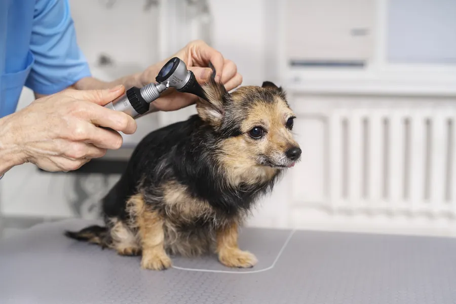
Your vet examines the ear canal and, where possible, the entire eardrum. The eardrum may be cloudy, thickened, bulging or ruptured. Video-otoscopy can provide a better view than conventional examination. A narrowed canal or one full of discharge may obscure the eardrum. Middle ear inflammation can be present behind an intact eardrum. In a study of dogs with longstanding outer ear inflammation, the eardrum was intact in 71.1% of ears with middle ear inflammation.
CT helps identify bony changes in the tympanic cavity wall. Contrast can help investigate a rupture if the eardrum cannot be seen during otoscopy. Contrast entered the tympanic cavity in 7 of 16 ears examined (44%), demonstrating a rupture. Five of these 7 ruptures were missed on video-otoscopy because the eardrum could not be seen adequately. MRI helps assess middle ear fluid and possible spread of inflammation into surrounding soft tissues. A brain MRI study found changes consistent with middle ear inflammation in 54 of 380 dogs (14.2%). Some findings were incidental, so imaging changes are assessed alongside symptoms. Middle ear fluid can appear on imaging without signs of ear canal inflammation. In the same 380-dog MRI study, 45 dogs had an inflammatory ear finding considered incidental (11.8%).
Sampling the middle ear
If the eardrum is intact and a middle ear sample is needed, your vet can make a small opening called a myringotomy. Sampling the middle ear matters because microbes in the canal may differ from those in the tympanic cavity. In one study, bacterial species or antimicrobial susceptibility differed between the canal and middle ear in 34 ears (89.5%). Canal material can contaminate a middle ear sample during collection, making culture harder to interpret. In one cadaver experiment, an ear canal marker reached the middle ear sample in 19 of 28 ears (67.9%). In another cadaver experiment using a new sampling technique, this occurred in 2 of 36 ears (5.55%). Microscopy can assess inflammatory cells in the sample. Bacterial culture and susceptibility testing help select an antimicrobial if disease-causing bacteria are identified.
Treatment
Flushing, medication and response
Your vet chooses treatment according to symptoms, eardrum condition and middle ear findings. The middle ear is flushed when needed to remove mucus and other material. Myringotomy and middle ear flushing are usually performed under general anaesthesia. A breathing tube protects the airway during flushing because fluid may drain through the auditory tube into the throat. Bacterial infection may be treated with topical and systemic antimicrobials. Pseudomonas infection can be difficult to treat because the bacterium can form a protective biofilm. Ear pain can be treated with pain relief prescribed by your vet. Some ear products can damage hearing if they enter the middle ear.
In a study of middle ear flushing and medication in 44 dogs, inflammation resolved in 36. The average time to resolution was 117 days. Times ranged from 30 to 360 days. Inflammation recurred in 7 of the 36 dogs in which it had resolved. Four dogs with recurrence needed further flushing. Of all 44 dogs, 4 eventually needed surgery. Follow-up information was unavailable for 3 dogs.
In secretory otitis media, a tenacious mucus plug is removed from the tympanic cavity if needed. A study describing 61 disease episodes in Cavalier King Charles Spaniels involved 43 dogs. Treatment in that study had to be repeated 1–5 times.
Surgery for severe or recurrent inflammation
For persistent inflammation unresponsive to other treatment, the tympanic cavity can be opened surgically. If the canal is permanently obstructed or calcified, it may also need removal. Opening the tympanic cavity aims to remove diseased tissue and allow drainage. When the ear canal is removed, the ear flap remains in place.
A study of subtotal ear canal removal and opening of the tympanic cavity included 18 French Bulldogs and 20 operated ears. Complete recovery was recorded in 9 of 18 dogs. Postoperative complications were recorded in 5 of 18 dogs. A mild head tilt present before surgery persisted in 5 of 18 dogs. Pre-existing facial nerve paralysis persisted in 4 of 18 dogs. One dog developed a deep infection after 4 months when a middle ear keratin mass recurred. Hearing loss may remain permanent even when the inflammation resolves.
Prevention
Treating outer ear infection and confirming recovery help reduce persistent inflammation. With recurrent outer ear inflammation, sustaining factors such as allergic skin disease are investigated. Regular ear checks help identify discharge and redness before a problem becomes longstanding. Drying the ears after swimming reduces moisture that may predispose dogs to outer ear infection. One Cavalier King Charles Spaniel breeding programme assesses secretory otitis media during head MRI. Under that breed programme, imaging is performed in dogs over 2 years old. A dog used for breeding must have the programme's required report before mating.
Important for dog owners
Record how long symptoms have been present, which ear has discharge and when it was last treated. Do not use drops left over from a previous ear infection on your own initiative. Avoid inserting a cotton bud into the canal because it can damage the eardrum. After surgery, a protective collar can prevent scratching of the wound. If your dog does not blink normally, your vet may advise lubricating drops to protect the eye's surface. Contact your practice about new discharge from the surgical area, as it may indicate residual infection. Any skin stitches are usually removed 10–14 days after surgery. A follow-up visit checks that ear inflammation has settled.
Frequently asked questions
Can a dog have middle ear inflammation with an intact eardrum?
Middle ear inflammation may lie behind an intact, normal-looking eardrum. With longstanding ear disease, your vet can assess the middle ear using CT or MRI. If a sample is needed, it can be collected through a small opening made in the eardrum.
Does fluid in the middle ear always mean bacterial infection?
Middle ear fluid may be an incidental finding on a short-muzzled dog's head MRI. Its significance is assessed using symptoms and other examination findings. Culture of middle ear fluid helps establish whether bacteria grow from that particular sample.
Can ear drops alone resolve middle ear infection?
Your vet plans treatment according to middle ear findings and the likely cause of inflammation. Thick material behind the eardrum may need removal by middle ear flushing under general anaesthesia. Bacterial infection may require systemic medication alongside topical treatment.
Can hearing return after treatment?
Hearing may deteriorate when middle ear fluid muffles sound transmission. It can improve as inflammation and fluid decrease. However, hearing loss may remain after severe or longstanding inflammation even when the ear is no longer inflamed.
What should I do if my dog develops a head tilt?
A head tilt may be associated with pain or a balance system disorder. Contact your vet the same day if head position changes suddenly or your dog starts walking unsteadily. If your dog can no longer remain standing, they need an emergency assessment.
Sources
- Mitä on päivystyshoito, Helsingin yliopisto, accessed 2026-11-07.
- Otitis Media and Interna in Animals, Merck Veterinary Manual, accessed 2026-11-07.
- Ulkokorvan tulehdus koirilla, Ruokavirasto, accessed 2026-11-07.
- Koiran korvatulehdus, Eläinklinikka Saari, accessed 2026-11-07.
- Prevalence of incidental and causative otitis in 380 client-owned dogs undergoing brain magnetic resonance imaging, Journal of Veterinary Internal Medicine, accessed 2026-11-07.
- Cytological and microbiological characteristics of middle ear effusions in brachycephalic dogs, Journal of Veterinary Internal Medicine, accessed 2026-11-07.
- Myringotomy in dogs: Contamination rate from the external ear canal, Veterinary Dermatology, accessed 2026-11-07.
- A novel video-endoscope-guided myringotomy technique in dogs, PeerJ, accessed 2026-11-07.
- Evaluation of canine tympanic membrane integrity using positive contrast computed tomography canalography, Frontiers in Veterinary Science, accessed 2026-11-07.
- Outcomes of subtotal ear canal ablation and lateral bulla osteotomy in French bulldogs, Canadian Veterinary Journal, accessed 2026-11-07.
- Otitis Externa, American College of Veterinary Surgeons, accessed 2026-11-07.
- Primary secretory otitis media in the Cavalier King Charles spaniel: a review of 61 cases, Journal of Small Animal Practice / Europe PMC, accessed 2026-11-07.
- Koiran perushoito, Suomen Kennelliitto, accessed 2026-11-07.
- Cavalier kingcharlesinspanielin jalostuksen tavoiteohjelma 2025–2029, Suomen Cavalier Kingcharlesinspanieliyhdistys, accessed 2026-11-07.
- Evaluation of outcome of otitis media after lavage of the tympanic bulla and long-term antimicrobial drug treatment in dogs: 44 cases (1998-2002)., Journal of the American Veterinary Medical Association / Europe PMC, accessed 2026-11-07.
- Microbial flora and antimicrobial susceptibility patterns of isolated pathogens from the horizontal ear canal and middle ear in dogs with otitis media., Journal of the American Veterinary Medical Association / Europe PMC, accessed 2026-11-07.
This information does not replace a visit to a vet. In an emergency, contact your nearest emergency vet: find a vet (RCVS).