
Dog diseases · Skin and coat
Nose and paw pad hyperkeratosis in dogs
Hyperkeratosis means a build-up of hard, dry, strand-like keratin on a dog's nose or paw pads. Changes in an ageing dog may cause no symptoms, but cracking or inflammation can make them painful. Inherited footpad disease in young dogs and hereditary nasal parakeratosis are separate diagnoses.
When to see a vet urgently
Pain, heavy bleeding or eye symptoms alongside thickened skin need urgent assessment:
- The pads have deep cracks and your dog limps or avoids walking.
- The nose bleeds heavily or repeatedly.
- Nasal ulcers occur alongside eye symptoms or deteriorating vision.
- Pad changes accompany widespread skin lesions and a clear deterioration in general health.
These findings may indicate a condition other than symptom-free keratin build-up. The vet guide can help you find emergency care.
Overview
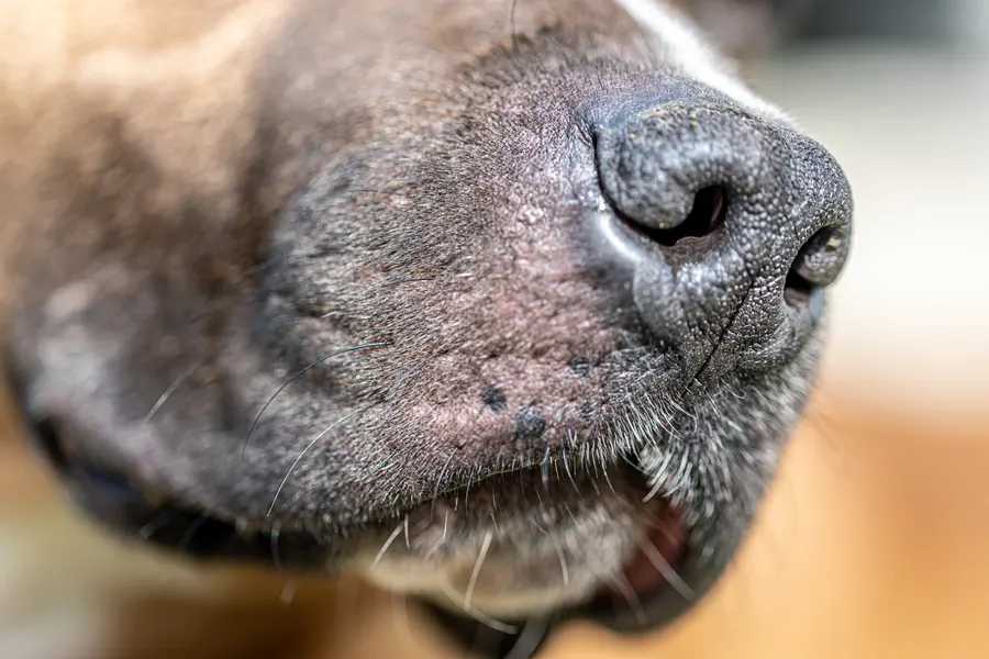
The nasal planum is the hairless nose surface where ordinary nasal hyperkeratosis mainly occurs. Dry keratin projections may grow particularly along the top edge of the nose or pad margins.
In the idiopathic form, no other underlying condition is identified and the dog is generally otherwise healthy. A US veterinary hospital study covering 1988–1998 identified 35 idiopathic cases.
These accounted for 0.4% of canine dermatology patients and 0.1% of all canine patients at the hospital. Only the nose was affected in 71.4%.
Both nose and pads were affected in 28.6%. These hospital figures describe the patients studied, rather than prevalence throughout other dog populations.
Causes
Keratinisation disorders are different conditions
In age-related idiopathic disease, excessive dry keratin gradually accumulates on the nose or pads. Hereditary footpad hyperkeratosis, or HFH, in Irish Terriers and Kromfohrlanders is associated with a FAM83G gene change.
This HFH form has autosomal recessive inheritance, so affected dogs receive variants from both parents. Hereditary nasal parakeratosis in Labrador Retrievers and Greyhounds involves abnormal cell maturation associated with SUV39H2.
The pad's outer keratin layer is normally specialised to withstand high mechanical loads during walking. In HFH, excessive surface thickening reduces flexibility and predisposes it to cracking.
The outermost layer thickens in HFH, but surface cells are mostly normally matured and have no visible nuclei in the keratin layer. In hereditary nasal parakeratosis, final cell maturation is disrupted, so similar appearances do not mean identical tissue changes.
Other conditions can also cause crusting
Zinc-responsive skin disease can cause nasal crusting and pigment loss. Hepatocutaneous syndrome, a liver-associated skin condition, can cause changes in the nose, pads and haired skin together.
Nasal crusting may also result from infection or immune-mediated skin disease.
Risk factors
Ordinary idiopathic nasal hyperkeratosis particularly affects older adult dogs. In the hospital study, age at onset was known for only nine of 35 dogs and ranged from four to nine years.
English Bulldogs had a relative risk of 14.3 in the hospital patient comparison. The corresponding figure for Miniature Poodles and Miniature Schnauzers was 9.5.
American Cocker Spaniels had a relative risk of 7.5. For Dobermanns, the figure was 2.6.
Zinc-responsive skin disease mainly affects northern breeds and often begins in young adulthood. The idiopathic hyperkeratosis study found no clear predisposition in either sex.
HFH may appear in Irish Terriers or Kromfohrlanders from about 4–5 months. Hereditary nasal parakeratosis in Labrador Retrievers may begin at about 6–12 months.
Typical symptoms
The pattern depends on whether there is keratin build-up alone or additional cracking and inflammation.
- Dry, hard or finger-like projections grow on the nose surface.
- An unusually thick keratin layer accumulates around the pad edges.
- The pad surface loses flexibility and cracks.
- Your dog avoids uneven ground or begins limping.
- Hereditary nasal parakeratosis may involve scaling, crusting and pigment loss.
- In hereditary footpad disease, nails may have a uniform thickness that makes trimming harder.
- With HFH, nails may also harden and appear to grow faster than usual.
- Some dogs with the hereditary footpad form also have abnormal coat quality.
In some cases, keratin build-up covers the whole nose or pad surface rather than just the edges. A young dog with hereditary nasal parakeratosis is also usually otherwise healthy, without signs of systemic disease.
Idiopathic nasal changes often mainly affect the top, with the nostrils and front surface remaining unchanged. Hereditary nasal parakeratosis can vary in severity over time, but changes are usually limited to the nose.
HFH may affect all pads rather than just one heavily loaded area.
Complications and consequences
Cracks change treatment needs
Ordinary nasal hyperkeratosis without inflammation may be completely painless. However, deep cracks can predispose the skin to secondary bacterial or Malassezia yeast infection.
Skin damage can cause pain and ulceration in previously symptom-free keratin build-up. Hereditary footpad hyperkeratosis may impair movement if it is not controlled.
Idiopathic changes do not usually disappear on their own, although they may remain similar for a long time. Long-term information was available for 25 of 35 dogs, so the later condition of every study dog was not known.
During hospital follow-up, changes remained stable for periods of one month to eight years. Hereditary nasal parakeratosis is lifelong, but generally has a good outlook with appropriate care.
Diagnosis at the vet
Examining the nose and all the skin
The assessment establishes age at onset and the original appearance, because crusting may hide the first sign. The vet also checks for similar changes elsewhere on the skin or pads.
Pigment loss, redness and loss of the normal cobblestone-like nasal surface may indicate inflammatory disease. With colour fading alone, surface structure may remain intact, as in vitiligo.
The ears are also examined, particularly with a dry nose, because ear inflammation can damage nerves involved in nasal secretion. A dry nose therefore does not automatically mean excessive keratin growth.
Samples and genetic tests
Cell samples can look for bacteria, yeasts and other cells associated with the lesion. Culture and susceptibility testing help select a suitable antimicrobial when needed.
Finding a microbe alone does not establish it as the original cause, because secondary overgrowth is common in an inflamed nose. A skin biopsy is useful when appearance alone cannot distinguish a keratinisation disorder from other nasal diseases.
Sampling aims to target an active, early lesion because an old ulcer may lose diagnostic tissue features. For an unclear nasal lesion, two small biopsies measuring 4–6 millimetres across may be taken, for example.
The sample must contain affected skin, because the surface layer may already be completely absent at the base of a deep ulcer. In certain inherited forms, a DNA test complements clinical assessment and tissue examination.
If hepatocutaneous syndrome is suspected, investigations may include abdominal ultrasound and blood and urine tests.
Treatment
Symptom-free changes and softening topical care
The vet prescribes treatment and any medication according to the cause, skin condition and other health issues. Symptom-free nasal hyperkeratosis in an older dog does not usually need medication for appearance alone.
If the keratin layer causes problems, treatment aims to soften it and remove excess keratin in a controlled way. A vet-selected petroleum jelly preparation or another suitable topical product may be used as an emollient.
Early softening in hereditary nasal parakeratosis may reduce deep cracking. Excess growth may need trimming after dry tissue has first been softened.
Using sharp tools or pulling off dry layers at home is not part of unsupervised care. The vet shows what can safely be done and which areas to leave alone. Treatment intensity is matched to severity, because a mild edge change and an extensively cracked surface need different approaches.
Infection and underlying disease need separate treatment
Bacterial infection may require a topical or systemic antimicrobial. Whenever possible, the choice is based on culture or other diagnostic findings.
An immune-mediated condition may require immune-suppressing treatment after diagnosis has been confirmed. Zinc treatment is considered for zinc-responsive skin disease, whose investigation also includes tissue examination.
A full response in confirmed zinc-responsive disease may take weeks or months. Softening keratin does not replace investigation and treatment of a cause such as hepatocutaneous syndrome.
Maintenance care for parakeratosis and idiopathic hyperkeratosis may be needed throughout life. Keratin growth may return after softening and trimming, so topical care continues according to the dog's needs.
Prevention
DNA testing can assess breeding-related risk for hereditary footpad hyperkeratosis. Some Irish Terrier show guidance asks judges to check the pads of at least one foot to detect abnormal keratin growth.
A 2022 Irish Terrier breed-club health survey identified one carrier through DNA testing. The footpad condition has recessive inheritance in Irish Terriers, so a parent without symptoms may carry the associated variant.
Hereditary nasal parakeratosis has a separate genetic test, because its gene change differs from FAM83G-associated footpad disease. Softening care can prevent severe cracking in existing hereditary nasal parakeratosis.
Important for dog owners
Take clear photographs of the nose and pads before trying topical products. Record age at onset and whether thickening, redness, fading colour or an ulcer appeared first.
Note whether all pads have changed and whether the paws are tender on different surfaces. Mention general condition, eating, drinking, eye and ear signs and other skin changes. Bring any DNA test result.
New lameness, pain or similar changes elsewhere on the skin are reasons to review the treatment plan.
Arrange a check if growth increases despite care. Ask the vet to show what safely treatable keratin looks like and how to recognise an ulcer beneath it.
Frequently asked questions
Does a dry nose always mean hyperkeratosis?
A dry nose can also involve nerve damage affecting nasal secretion, caused by an ear infection, for example. Hyperkeratosis involves visible keratin growth on the nose. Examining the ears may therefore form part of investigating dryness. An unclear skin lesion may need a biopsy to distinguish other nasal conditions.
Can symptom-free nasal keratin build-up be harmless?
Non-inflammatory nasal hyperkeratosis in an older adult dog may be painless and usually needs no treatment. Cracking or inflammation changes the situation, and topical care may become necessary. Deep fissures can predispose the skin to bacterial or yeast overgrowth.
Why are my puppy's pads hardening and cracking?
In Irish Terriers and Kromfohrlanders, the first signs of hereditary footpad hyperkeratosis may appear at 18–24 weeks. The thickened pad surface loses flexibility and cracks. A DNA test is available to investigate the inherited form.
Can I cut the thickened surface of the nose myself?
Do not start cutting or pulling it away yourself; agree a method with the vet. Treatment uses softening of dry keratin and trimming of excess growth when necessary. A cracked or ulcerated surface may be painful and needs appropriate care.
Does moisturising cure inherited disease?
Hereditary nasal parakeratosis is permanent, and topical care does not provide a lasting cure. Nevertheless, softening can reduce severe cracking and help maintain a healthier surface. The outlook is generally good despite the need for long-term care.
Sources
- Nasal Dermatoses of Dogs, Merck Veterinary Manual, accessed 2026-11-03.
- Idiopathic Nasodigital Hyperkeratosis in Dogs: A Retrospective Analysis of 35 Cases (1988–1998), Japanese Journal of Veterinary Dermatology, accessed 2026-11-03.
- Irlanninterrierin jalostuksen tavoiteohjelma 2025–2029, Suomen Irlanninterrierit ry, accessed 2026-11-03.
- Nasal planum diseases in dogs, Canadian Veterinary Journal May 2024, Canadian Veterinary Medical Association, accessed 2026-11-03.
- A Mutation in the FAM83G Gene in Dogs with Hereditary Footpad Hyperkeratosis (HFH), PLOS Genetics, accessed 2026-11-03.
- Conditions Affecting the Nasal Planum in Canine Patients, Clinician’s Brief, June 2018, accessed 2026-11-03.
- Veterinary Medicine Community Client Instructions – Nasodigital Hyperkeratosis, Elsevier veterinary client education, accessed 2026-11-03.
This information does not replace a visit to a vet. In an emergency, contact your nearest emergency vet: find a vet (RCVS).
More on this topic: Skin and coat
- Acne in dogs: chin spots and treatment
- Alopecia X and seasonal flank alopecia in dogs
- Paw pad injuries in dogs: first aid and wound care
- Atopic dermatitis in dogs: itching, tests and long-term care
- Demodectic mange in dogs: Demodex mites and skin disease
- Fibrosarcoma in dogs: symptoms, diagnosis and treatment