
Dog diseases · Eyes, ears and mouth
Oral squamous cell carcinoma in dogs
Oral squamous cell carcinoma is a malignant tumour found in sites including the gums, beneath the tongue and the tonsils. It may appear as a bleeding lump or ulcer. The tumour type is established from a biopsy.
When to see a vet urgently
Arrange a veterinary appointment soon if your dog has a lump or ulcer in the mouth that does not heal. Emergency care is needed if any of the following signs occur.
- Your dog has laboured breathing.
- The mouth bleeds heavily or bleeding will not stop.
- Your dog cannot swallow water.
- Your dog has severe mouth pain.
- Their gums become pale or they cannot remain standing.
Overview
Tumour location and type
Squamous cell carcinoma originates in the lining of the mouth. It is the second most common malignant oral tumour in dogs. At one veterinary hospital, 79 of the oral tumours in 526 dogs were squamous cell carcinomas (15.02%). In that study, 59 of the 79 carcinomas were on the gums (74.68%). Eight were in a tonsil (10.13%). Forms growing beneath the tongue or in a tonsil metastasise more readily than tumours at the front of the mouth. There is also a rare papillary form, in which surface cells form projections. In this form, cancer cells can grow into supporting tissue beneath the epithelium despite wart-like projections on the surface. Appearance alone may make papillary carcinoma difficult to distinguish from a benign oral wart.
Causes
No single known cause of canine oral squamous cell carcinoma has been identified. Changes in the activity of genes controlling cell division have been found in tumour tissue. Increased activity of the growth-regulating RAS signalling pathway has been identified in tumour cells. Reduced activity of genes involved in normal oral epithelial differentiation has also been observed. Cancer cells can invade tissue beneath the surface lining and then the jawbone.
One study found canine papillomavirus genetic material in 3 of 33 oral squamous cell carcinoma tissue samples. Active viral gene transcription could not be demonstrated in these carcinomas. A persistent viral oral wart can become cancerous, but this is rare. In one case series, papillomas in 7 dogs showed tissue changes consistent with malignant transformation.
Risk factors
Conventional oral squamous cell carcinoma is diagnosed more often in older dogs. Among 40 gingival carcinoma patients treated with jaw surgery, the median age at diagnosis was 9.5 years. Ages in that group ranged from 0.5 to 15.5 years. No individual breed predisposition was identified in this gingival carcinoma group. The tumour has been reported more often in breeds such as Cocker Spaniels, retrievers and Poodles, but a definite breed predisposition is not established.
The papillary form can develop in puppies. However, it has also been diagnosed in adult and older dogs. Ten dogs selected for radiotherapy for papillary carcinoma had a median age of 4 years when treatment began. Four were under 1 year old.
Typical symptoms
An oral tumour may cause no symptoms initially.
- A reddish lump or thickened ulcer may be visible on the gum or elsewhere in the mouth.
- Your dog may drool more, and the saliva may contain blood.
- Their breath may smell unpleasant.
- Food may fall from the mouth or chewing may become difficult.
- Opening the mouth may be painful.
- Teeth beside the tumour may loosen.
- One side of the muzzle or jaw may swell.
A tonsillar tumour can be difficult to see at the back of the mouth. The first finding may therefore be a lump in the neck caused by an enlarged lymph node. Persistent eating difficulties may lead to weight loss.
Complications and consequences
Local tissue damage and metastases
The visible lump may be smaller than the part growing deep within the tissues. A tumour invading the jawbone can cause pain without a large visible ulcer. Rarely, the jawbone can fracture where the tumour has weakened it. A local tumour may be accompanied by bacterial infection. A tumour in the upper jaw can extend into the nasal cavity. Sneezing or nasal discharge may accompany an upper jaw lesion if the tumour extends into the nasal region.
A tonsillar carcinoma can spread to mandibular or retropharyngeal lymph nodes. The lungs are one possible site of distant metastases. In the surgical study of 40 dogs with gingival carcinoma, 2 had lymph node metastases at initial assessment (5%). In another study, metastases were found in 6 of 21 dogs with oral squamous cell carcinoma (29%) when lymph nodes were removed for tissue examination.
Diagnosis at the vet
Oral examination, tissue sampling and imaging
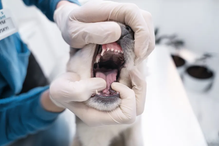
Your vet examines the whole mouth because a tumour's edge or a lesion beneath the tongue can be difficult to see in an awake dog. Sedation may be needed for a thorough oral examination. An early tumour can resemble inflammation of the tooth-supporting tissues. Tissue appearance cannot establish whether a lump is benign or malignant. A pathologist examines a tumour biopsy under a microscope. Tissue sampling distinguishes wart-like papillary carcinoma from a benign papilloma. Inflammation and dead tissue can make a fine-needle sample difficult to interpret. A representative tissue sample may then be taken from within the lesion, for example as a wedge biopsy. Dental X-rays can show whether the lesion extends into surrounding bone. CT helps assess the extent of bone damage before surgery.
Assessing spread
Your vet may take a fine-needle sample from a regional lymph node to look for metastases. Lymph from the mouth can drain into mandibular lymph nodes on both sides. An enlarged lymph node can also be reacting to inflammation. In a study of malignant oral tumours, 4 of 17 dogs had metastases on the side opposite the tumour when lymph nodes on that side were also removed. Chest imaging looks for spread to the lungs. TNM staging assesses the extent of the primary tumour, regional lymph nodes and distant metastases. T1 means a primary tumour under 2 cm in diameter. A tumour measuring 2–4 cm is classified as T2. A tumour over 4 cm is classified as T3. Bone involvement is recorded separately because tumours of the same size can extend to different depths.
Treatment
Surgery and assessment of margins
Your vet chooses treatment according to the tumour's size, location and extent of spread. First-line treatment for a localised tumour is usually surgery to remove it with sufficiently wide margins. If it has invaded the jawbone, part of the jaw may need to be removed with it. A tumour arising in the tongue can be treated by removing part of the tongue. The edges of the surgical specimen are examined to check for healthy tissue surrounding the tumour. If tumour cells are present at an edge, the risk of local recurrence is greater.
The 40 gingival carcinoma patients treated with jaw surgery had a median survival of 44.8 months. The extent of spread was associated with survival in this group. Of 97 dogs undergoing tongue tumour surgery, 31 had squamous cell carcinoma (32%). Dogs with squamous cell carcinoma of the tongue had a median survival of 216 days. A tongue tumour diameter of at least 2 cm was associated with shorter survival. Across all tumour types in the tongue surgery series, local recurrence occurred in 27 dogs (28%).
Radiotherapy and symptom relief
Radiotherapy may be used after surgery if removal was incomplete. A study assessing surgery combined with radiotherapy included 87 dogs with either non-tonsillar squamous cell carcinoma or fibrosarcoma. In the incompletely excised squamous cell carcinoma group, median survival without radiotherapy was 181 days. Median survival was substantially longer in the radiotherapy group of the same study. If a tumour cannot be operated on, radiotherapy can be used to relieve symptoms. In 10 of 13 dogs with gingival carcinoma (77%), the visible tumour shrank or disappeared after palliative radiotherapy. Median survival in that group was 365 days.
In another study, symptoms improved after stereotactic radiotherapy in 17 of 18 dogs with oral squamous cell carcinoma (94.4%). Median survival in that study's carcinoma group was 332 days. Bone necrosis or a fistula occurred in 9 of these 18 dogs (50%). In a gingival carcinoma radiotherapy study, oral mucositis occurred in 9 of 21 dogs. Late radiation effects occurred in 3 palliatively treated dogs, representing 14% of all 21 dogs.
Among 10 dogs with papillary carcinoma, the visible tumour disappeared after radiotherapy in 9. In the only dog with a partial response, the tumour progressed again 228 days after radiotherapy began. Pain relief and nutritional support form part of symptomatic care. Antibiotics are used when needed for bacterial infection associated with the tumour.
Outlook for tonsillar carcinoma
Among 15 dogs with tonsillar squamous cell carcinoma, median survival was 243 days. When the tumour was confined to 1 tonsil without identified metastases, median survival was 637.5 days. With local metastases, it was 134 days. Dogs with distant metastases had a median survival of 75 days. In the same study, 40% of dogs were alive at 1 year. At 2 years, 20% were alive. In a larger study of different tonsillar carcinomas, median survival among 123 dogs was 126 days.
Recovery after jaw surgery
After upper jaw surgery, a fistula may remain between the mouth and nasal cavity. After lower jaw surgery, the lower jaw may shift sideways and the bite may change. In a study of jaw surgery in 459 dogs, 171 had at least one complication (37.3%). Of 253 complications graded for severity, 157 were minor (62.1%). Of 271 recorded complications, 90.0% occurred within 3 months of surgery. In another study, 71% of responding owners reported that their dog took food and water normally within 4 weeks of jaw surgery.
Prevention
There is no known specific way to prevent oral squamous cell carcinoma. Regular oral examinations may allow your vet to notice a tumour before symptoms develop. A small tongue tumour can be detected by examining the upper and lower surfaces before swallowing becomes difficult. In one study of several oral tumour types, 15 tumours were found incidentally during dental treatment (15.3% of 98 dogs). Regular toothbrushing helps prevent tartar formation and provides an opportunity to notice a new gum lesion. Introducing toothbrushing in puppyhood makes regular brushing easier.
Important for dog owners
Record when you noticed the lesion and whether it has grown or started bleeding. Watch for food left in the bowl or dropped from the mouth. After jaw surgery, your dog may need soft food for 2–3 weeks as your vet advises. Hard treats and chew toys can rub the mouth while the surgical wound heals. Contact your practice if the surgical area swells or the wound opens. Follow-up visits check for new growth at the surgical site. After extensive surgery to the front of the lower jaw, the tongue may remain visible and drooling may increase.
Frequently asked questions
Does a lump on the gum mean cancer?
Oral lumps can be benign or malignant. A fistula associated with a tooth root infection can also cause swelling resembling a tumour. If the cause is unclear, examining a biopsy helps distinguish a tumour from other oral lesions.
Why might even a small lump need a CT scan?
A lesion visible on the mouth's surface can be smaller than the tumour invading deeper tissues. CT shows how far the tumour has progressed in bone. The images help plan surgery with the most appropriate possible removal margins.
Can a dog eat after part of the jaw is removed?
Many dogs adapt to eating after removal of part of the jaw. However, food may fall from the mouth more easily, particularly after extensive upper jaw surgery. During recovery, your vet may advise soft food that your dog can lap into their mouth.
Why is tonsillar carcinoma discussed separately?
Tonsillar squamous cell carcinoma spreads to lymph nodes more readily than the form growing at the front of the mouth. Metastases may be the first finding when the tonsillar lesion is small. Assessing spread helps estimate the outlook and choose suitable treatment for your dog.
Can the tumour be treated without surgery?
Radiotherapy is one option if the tumour cannot be removed with sufficiently wide margins. Palliative radiotherapy may ease pain and reduce bleeding from the visible tumour. If neither treatment is suitable, your vet can support eating and relieve pain.
Sources
- Mitä on päivystyshoito, Helsingin yliopisto, accessed 2026-11-07.
- Suun kasvaimet koirilla ja kissoilla, Eläinklinikka Tsemppi, accessed 2026-11-07.
- Oral squamous cell carcinoma in dogs, Cornell University College of Veterinary Medicine, accessed 2026-11-07.
- Oral Tumors, American College of Veterinary Surgeons, accessed 2026-11-07.
- A possible role of coarse fractionated radiotherapy in the management of gingival squamous cell carcinoma in dogs, Veterinary Medicine and Science, accessed 2026-11-07.
- Tonsillar carcinoma in dogs: Treatment outcome and potential prognostic factors in 123 cases, Journal of Veterinary Internal Medicine, accessed 2026-11-07.
- Treatment of canine oral papillary squamous cell carcinoma using definitive-intent radiation as a monotherapy, Veterinary Medicine and Science, accessed 2026-11-07.
- Outcomes following surgical excision or surgical excision combined with adjunctive, hypofractionated radiotherapy in dogs with oral squamous cell carcinoma or fibrosarcoma, University of Cambridge / JAVMA, accessed 2026-11-07.
- Characteristics of canine oral tumors: Insights into prevalence, types, and lesion distribution, Open Veterinary Journal, accessed 2026-11-07.
- A Retrospective Investigation on Canine Papillomavirus 1 in Oral Oncogenesis, PLOS ONE, accessed 2026-11-07.
- Malignant transformation of canine oral papillomavirus-associated papillomas in dogs, Papillomavirus Research, accessed 2026-11-07.
- Pathology in Practice: Canine oral papillomas and squamous cell carcinoma, JAVMA, accessed 2026-11-07.
- Radiation therapy of canine nontonsillar squamous cell carcinoma, Canadian Veterinary Journal, accessed 2026-11-07.
- Sisätaudit: kasvainsairaudet, Helsingin yliopisto, accessed 2026-11-07.
- Quality of Life After Partial Mandibulectomy or Maxillectomy in 45 Dogs With Oral Tumors., Journal of veterinary dentistry / Europe PMC, accessed 2026-11-07.
- Analysis of risk factors associated with complications following mandibulectomy and maxillectomy in dogs., Journal of the American Veterinary Medical Association / Europe PMC, accessed 2026-11-07.
- Evaluation of the factors contributing to long-term survival in canine tonsillar squamous cell carcinoma., Australian veterinary journal / Europe PMC, accessed 2026-11-07.
- Response of Spontaneous Oral Tumors in Canine Cancer Patients Treated with Stereotactic Body Radiation Therapy, Radiation Research, accessed 2026-11-07.
- Conference 23, Case 04: Papillary squamous cell carcinoma, Joint Pathology Center, accessed 2026-11-07.
- Mandibulectomy or Maxillectomy, Southwest Veterinary Surgical Service, accessed 2026-11-07.
- Results of surgical excision and evaluation of factors associated with survival time in dogs with lingual neoplasia: 97 cases (1995-2008)., Journal of the American Veterinary Medical Association / Europe PMC, accessed 2026-11-07.
- [Prognosis of canine oral (gingival) squamous cell carcinoma after surgical therapy. A retrospective analysis in 40 patients]., Tierarztliche Praxis. Ausgabe K, Kleintiere/Heimtiere / Europe PMC, accessed 2026-11-07.
- Molecular homology between canine spontaneous oral squamous cell carcinomas and human head-and-neck squamous cell carcinomas, Neoplasia, accessed 2026-11-07.
- Oral Masses in Dogs and Cats, Delaware Veterinary Dental Practice, accessed 2026-11-07.
- Koiran perushoito, Suomen Kennelliitto, accessed 2026-11-07.
- Surgical Options for Oral Tumors, MSPCA-Angell, accessed 2026-11-07.
- Histologic evaluation of mandibular and medial retropharyngeal lymph nodes during staging of oral malignant melanoma and squamous cell carcinoma in dogs, JAVMA / University of Minnesota, accessed 2026-11-07.
- The MEK inhibitor trametinib is effective in inhibiting the growth of canine oral squamous cell carcinoma, Scientific Reports, accessed 2026-11-07.
This information does not replace a visit to a vet. In an emergency, contact your nearest emergency vet: find a vet (RCVS).