
Dog diseases · Eyes, ears and mouth
Progressive retinal atrophy in dogs
Progressive retinal atrophy, or PRA, is a group of inherited diseases gradually damaging light-sensitive cells. Vision loss often first appears in dim light. Progression is usually slow, and PRA itself is painless.
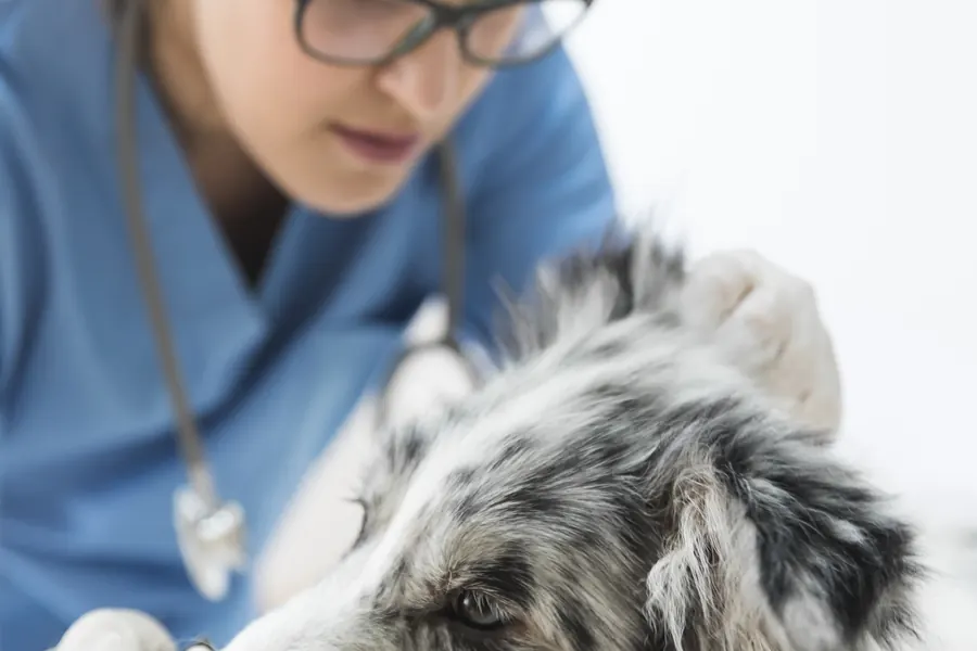
When to see a vet urgently
Arrange an appointment within days for hesitation in dim light or collisions in unfamiliar surroundings. Contact an emergency clinic promptly for these changes.
- Sudden blindness or collisions with familiar obstacles.
- Severe squinting or holding an eye closed through pain.
- Sudden surface cloudiness.
- Altered shape or bulging.
- Serious eye injury.
Overview
How the retina detects light
The retina lies at the back of the eye. The lens focuses light onto it. Photoreceptors convert light into electrical signals sent to the brain. Rods support dim-light vision and movement detection. Cones support daylight and colour vision. Dogs have more rods than cones. Most PRA forms affect rods first. Cone damage then impairs vision in bright surroundings too. Cell loss is usually bilateral and fairly symmetrical. Mild congenital retinal folds are generally stable, whereas PRA progressively reduces vision.
Causes
Forms and inheritance
Several genetic variants can cause PRA. More than one form can occur within a breed. Late-onset forms initially develop normal photoreceptors. Early inherited disorders can disrupt their development in puppies. Such developmental abnormalities may be termed retinal dysplasia.
Most PRA is autosomal recessive. At-risk dogs inherit the associated variant from both parents. Some dominant forms need only one copy. X-linked forms affect males more often. RPGR-associated X-linked PRA has been described in Siberian Huskies. Irish Setter rcd1 rapidly destroys photoreceptors. It involves PDE6B, important for rod light responses. Late-onset rcd4 occurs in Gordon and Irish Setters, among others.
Risk factors
Purebred and crossbred dogs can be affected. Associated variants occur in breeds such as Poodles, Labrador Retrievers and Cocker Spaniels. Golden Retrievers have inherited retinal degenerations beyond prcd-PRA.
Lapponian Herders have PRCD- and IFT122-associated forms. A gene bank study from northern Europe found IFT122 carriers among 28% of 519 examined Lapponian Herders. Among 577 Finnish Lapphunds, 12% were carriers. The variant's visual significance in Finnish Lapphunds needs further study. All 10 affected Lapponian Herders in the initial comparison had two copies. Among the same 577 Finnish Lapphunds, seven PRA patients unexplained by PRCD lacked IFT122 variants too.
Several associated forms occur in Miniature Schnauzers. HIVEP3 and PPT1 variants in the same chromosomal region are being investigated for one form. HIVEP3 can serve as a risk-linked marker in testing. If marker and causal variant are not inherited together, results can mislead. Among 51 affected Miniature Schnauzers, average diagnosis age was 3.9 years.
One breed health report from northern Europe recorded PRA fundus changes in 14 Spanish Water Dogs by the end of 2020.
Typical symptoms
Dim-light vision can decline before noticeable daytime changes. Familiar home navigation can remain normal despite difficulties elsewhere.
- Hesitation entering dark rooms.
- Slowing or stopping at the top of stairs.
- Colliding with doorframes in unfamiliar places.
- Larger-looking pupils.
- Brighter reflected light from the eyes.
- Later lens cloudiness from cataracts.
Adult-onset PRA is often diagnosed at three to nine years. Early developmental photoreceptor disorders can be diagnosed around two to three months. Onset varies within breeds and between forms. IFT122-associated PRA was diagnosed at an average nine years in Lapponian Herders. One affected 13-year-old retained some vision.
Complications and consequences
Declining vision and secondary cataracts
Both eyes usually lose vision. Most affected dogs eventually become completely blind. This can take one to two years, but progression depends on form and individual.
Secondary cataracts can obscure the retina during fundus examination. Leaking lens material can inflame the interior. This can hurt despite painless PRA.
Diagnosis at the vet
Fundus examination and retinal electrical testing
The vet establishes which lighting first caused difficulty. Pupil light responses are assessed. Constriction may be slow. Thinning can increase fundus reflectivity. Retinal vessels can look narrower. Advanced disease can also atrophy the optic disc. Early degeneration may be invisible on routine examination.
Electroretinography, or ERG, measures function. It can detect dysfunction before visible degeneration. It also works when cataracts obscure the retina. Optical coherence tomography, or OCT, measures layer thickness. It can monitor structural change over time. Dark- and light-adapted ERG separately assesses rods and cones.
Progression and other vision-loss causes
Four prcd-PRA research dogs had normal-looking reflective fundus areas until age 2.3 years. Rod responses were reduced versus controls by 0.5 years. The photoreceptor nuclear layer was thinned in every examined quadrant by 0.8 years. Cone responses were better preserved.
OCT measured 113 µm retinal thickness in a Lapponian Herder with two IFT122 copies. A healthy control measured 185 µm. This comparison involved one affected and one healthy Lapponian Herder.
Sudden acquired retinal degeneration syndrome, or SARDS, can cause blindness within days or weeks. ERG responses can be absent despite initially normal fundus appearance. Retinal detachment treatment targets its cause. Acute glaucoma can also rapidly destroy vision.
Genetic test information
Blood or cheek cells can be tested for a specific variant. Testing can identify asymptomatic recessive carriers. Not every PRA variant is known. One negative test cannot exclude another genetic form. Eye examination assesses disease signs despite a known variant. Lapponian Herder prcd-PRA and IFT122 tests can be performed at any age.
Treatment
Monitoring vision and other eye disease
No curative treatment is available for PRA. Daily support aims to reduce injuries as sight declines. Secondary cataract-related internal inflammation is assessed separately. Treatment is prescribed according to examination findings. Cataract surgery cannot restore function lost through a non-functioning retina. Antioxidant supplements have no demonstrated effect on PRA progression. Some have been proposed to slow cataract formation. Ultrasound before possible surgery can check retinal attachment.
Gene therapy research
Experimental therapy targets a specific causal gene. RPGR-targeted treatment preserved retinal cells in dogs with X-linked degeneration. Visual function persisted for over two years. Benefits can be limited to treated retinal areas. Success requires remaining retinal structure. Canine PRA gene therapy remains a research treatment.
Prevention
Some kennel club breeding guidance excludes affected dogs. Recessive carriers can be paired with dogs clear of the same variant. Two prcd-PRA carriers produce an average 25% offspring inheriting both copies. An average 50% are carriers. An average 25% inherit neither copy.
Some Lapponian Herder breeding guidance separately considers prcd-PRA and IFT122-PRA. Some breed health programmes require eye examinations before breeding. Some official registries require genetic samples collected by a vet or approved sampler. Their official results require sampling details on an electronic referral. Results can guide mate selection while preserving genetic diversity. The prcd-PRA variant declined in tested Labrador Retrievers during 2004–2019.
Important for dog owners
Improve route lighting for dim-light hesitation. Keep furniture in familiar positions to support navigation from memory. Gate off open stairs. Use a lead in unfamiliar places. Teach verbal cues before sight is lost. Smell and hearing can support navigation. Daylight walks can help route-finding after night vision declines. Fenced gardens can provide safe off-lead space.
Frequently asked questions
Can affected dogs see during the day?
Early in the disease, a dog may still see in daylight even though dim-light vision has already deteriorated. Daytime vision declines when the cone cells in the retina are also damaged. The first noticeable difficulties may occur somewhere unfamiliar, where your dog does not already know the routes.
Can a carrier see normally?
A dog carrying one copy of the recessive prcd-PRA variant can have normal vision. A carrier can nevertheless pass the variant on to a puppy. When planning breeding, it is therefore important to check that the proposed mate does not carry the same recessive variant. The genetic result shows which variant the dog carries and can pass to offspring.
Does a clear genetic result rule out PRA?
A result provides information only about the variants included in that particular test. Several different forms of PRA can occur within the same breed. If your dog loses vision, the vet examines the eyes and can assess retinal function with ERG when needed.
Can cataract surgery restore sight with PRA?
PRA reduces the ability of the retina to detect light. During cataract surgery, the cloudy lens is removed from the eye. Before considering surgery, the vet can use ERG to assess whether sufficient retinal function remains. Removing lens opacity cannot restore sight if retinal function is lost.
How do dogs cope with declining sight?
Many dogs adapt well to a slowly progressing loss of vision. Keeping familiar routes makes it easier for your dog to move around at home. Your dog can use smell and hearing to find the way in familiar surroundings. Protecting stairs with a safety gate prevents your dog falling from the top.
Sources
- Silmäsairaudet ja -tutkimukset, Suomen Kennelliitto, accessed 2026-11-07.
- Silmäsairaudet, Helsingin yliopisto, accessed 2026-11-07.
- Overview of Ophthalmic Emergencies in Small Animals, Merck Veterinary Manual, accessed 2026-11-07.
- Progressive retinal atrophy, Cornell University, accessed 2026-11-07.
- Progressive Retinal Atrophy in Dogs, VCA Animal Hospitals, accessed 2026-11-07.
- Espanjanvesikoiran jalostuksen tavoiteohjelma 2022–2026, Vesikoirat ry, accessed 2026-11-07.
- Consensus guidelines for nomenclature of companion animal inherited retinal disorders, Veterinary Ophthalmology, accessed 2026-11-07.
- Koiratutkimus paljasti uuden sokeuteen vaikuttavan geenin, Helsingin yliopisto, accessed 2026-11-07.
- A missense variant in IFT122 associated with a canine model of retinitis pigmentosa, Human Genetics, accessed 2026-11-07.
- PRCD-associated retinitis pigmentosa in dogs and humans, Experimental Eye Research, accessed 2026-11-07.
- Lapinporokoirien jalostuskriteerit, Lappalaiskoirat ry, accessed 2026-11-07.
- Ohjeet terveystutkimusten ja geenitestien teettämiseen, Lappalaiskoirat ry, accessed 2026-11-07.
- Kääpiösnautseri, Suomen Kennelliitto, accessed 2026-11-07.
- Complex Structural PPT1 Variant Associated with Non-syndromic Canine Retinal Degeneration, G3: Genes, Genomes, Genetics, accessed 2026-11-07.
- Global Frequency Analyses of Canine Progressive Rod-Cone Degeneration–Progressive Retinal Atrophy and Collie Eye Anomaly, Genes, accessed 2026-11-07.
- Successful arrest of photoreceptor and vision loss expands the therapeutic window of retinal gene therapy, Proceedings of the National Academy of Sciences, accessed 2026-11-07.
- Sudden Acquired Retinal Degeneration Syndrome (SARDS), VCA Animal Hospitals, accessed 2026-11-07.
- The genetics of inherited retinal disorders in dogs: implications for diagnosis and management, Veterinary Medicine: Research and Reports, accessed 2026-11-07.
- My dog is going blind due to progressive retinal atrophy - is gene therapy an option?, American College of Veterinary Ophthalmologists, accessed 2026-11-07.
- Kultainennoutaja, Suomen Kennelliitto, accessed 2026-11-07.
- Joint Formal Comment for PLGE and Commentary for G3, PLOS Genetics, accessed 2026-11-07.
- Geenitestit, Suomen Kennelliitto, accessed 2026-11-07.
- The Lens in Animals, Merck Veterinary Manual, accessed 2026-11-07.
- Cataract Surgery, University of Florida College of Veterinary Medicine, accessed 2026-11-07.
This information does not replace a visit to a vet. In an emergency, contact your nearest emergency vet: find a vet (RCVS).