
Dog diseases · Digestive system
Pulmonary oedema in dogs: symptoms and emergency treatment
Pulmonary oedema means fluid accumulating in the lung tissue and air sacs. Breathing becomes faster and more difficult. Heart failure or disrupted lung fluid balance without heart disease may cause it. Breathing difficulty is an emergency.
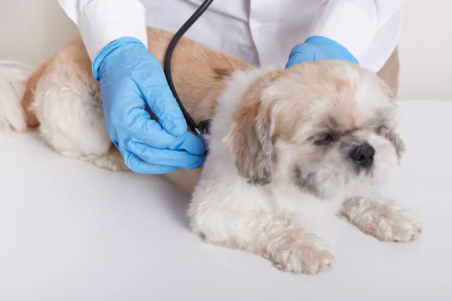
When to see a vet urgently
A dog struggling to breathe needs immediate help, because severe oxygen deprivation can lead to respiratory arrest.
- Breathing at rest is clearly harder or faster than usual.
- Your dog cannot lie down and instead sits or stands with the neck extended.
- The gums or tongue look blue or grey during breathing difficulty.
- Your dog breathes with the mouth open and appears to struggle for air.
- Your dog collapses, cannot stand or becomes less conscious.
Overview
Pulmonary oedema results from various diseases, so treatment also requires investigation of the fluid accumulation's cause. Cardiogenic means heart-related, while non-cardiogenic means unrelated to the heart. Both forms may require supplemental oxygen to maintain oxygen delivery.
Capillaries surround the air sacs, where gases are exchanged between blood and breathed air. Lymphatic vessels normally remove fluid seeping into lung tissue. Pulmonary oedema develops when fluid accumulates faster than it can be removed.
It differs from pleural effusion, where fluid lies outside the lungs in the chest cavity. Pneumonia can cause similar breathing difficulty, but involves inflammation or infection in lung tissue.
Causes
Heart-related disease raises pressure in the pulmonary circulation
Left-sided heart failure can raise venous pressure enough for fluid to move into the lungs. A leaking mitral valve or heart muscle disease may underlie it. The body may retain salt and water, increasing blood volume and further straining the heart. Rupture of a valve-supporting cord can cause sudden cardiogenic pulmonary oedema.
There are several non-cardiogenic causes
Other causes include head injury, severe seizures, electric shock and airway obstruction. Breathing against a blocked airway creates strong negative pressure in the chest. This changes lung pressure differences and can draw fluid from capillaries into air sacs.
Pulmonary oedema can also develop during treatment because of excessive fluid loading. A rare separate form involves re-expansion of a previously collapsed lung, for example after chest surgery.
Inflammation can increase permeability of the lung's blood–air barrier, allowing fluid into lung tissue more readily. Drowning or a severe inflammatory response can also cause non-cardiogenic pulmonary oedema. ARDS is acute respiratory distress syndrome, an inflammatory lung condition that can result from disease in the lungs or elsewhere. Hunting-associated pulmonary oedema is also recognised in Drevers, although its precise mechanism remains unclear.
Risk factors
Cardiogenic risk depends on the heart disease present and how far it has progressed. Degenerative mitral valve disease is common particularly in older small and medium-sized dogs. Predisposed breeds include Cavalier King Charles Spaniels and Dachshunds. Valve disease alone does not mean pulmonary oedema: around 25–50% develop signs of clinical heart failure.
Airway obstruction, chewing an electrical cable or head injury predisposes through a different mechanism from heart disease. A post-hunting form has been described in Drevers and Dachshunds. In one Drever symptom questionnaire from northern Europe, median onset age was 29.8 months in males and 43.3 months in females. These figures describe dogs with signs rather than the risk for the entire Drever population.
Typical symptoms
A rising respiratory rate can be the first sign of heart-related fluid accumulation before visible breathing difficulty. You may notice the following changes:
- Breathing at rest or during sleep becomes faster than before.
- Breathing looks laboured and deeper than usual.
- Your dog stands or sits because lying down makes breathing difficult.
- Your dog extends the neck and holds the elbows away from the body.
- Your dog coughs or tolerates exercise less well.
- In severe cases, the mouth is open and mucous membranes look blue.
Coughing can accompany pulmonary oedema, but airway disease is often its cause in dogs. A cough together with a heart murmur therefore does not confirm heart failure.
Complications and consequences
Severe pulmonary oedema can cause respiratory failure and death. The outlook depends on lung injury severity and the disease responsible. ARDS is particularly serious: canine mortality in two retrospective studies was 84% and 92%. These figures concern ARDS patients rather than all dogs with pulmonary oedema.
After heart failure begins due to degenerative mitral valve disease, many treated dogs live around another 12–18 months. This estimate concerns valve disease rather than lung injury after electric shock or airway obstruction. Hunting-associated oedema in Drevers can recur: 52.9% of dogs in the symptom questionnaire had experienced at least 5 episodes.
Diagnosis at the vet
Breathing safety is assessed first
At presentation, breathing effort, mucous membrane colour and posture are assessed before stressful investigations. Lung crackles may be heard, but breath sounds can also be normal. Oxygenation is monitored with pulse oximetry or arterial blood gas measurement. Stabilisation begins before extensive imaging in a dog with dangerous breathing difficulty.
Important history includes heart disease and any seizure, choking, electric shock or immersion in water. In hunting dogs, the relationship between breathing signs and pursuit helps assess hunting-associated oedema.
Imaging examines both lungs and heart
Chest X-rays look for lung fluid and changes in the heart or pulmonary vessels. Echocardiography shows changes in valves, heart chambers and pumping function. Point-of-care lung ultrasound can help rapid assessment and response monitoring.
Lung ultrasound is generally performed standing or lying on the chest, allowing both sides to be assessed without changes caused by lying on one side. Shaving is usually unnecessary, although local clipping may be needed if the coat prevents adequate imaging. The examination is external, and a portable machine produces real-time images. It can also look for pleural fluid, pneumothorax or other lung tissue changes. Ultrasound B-lines indicate altered lung tissue but can also occur with conditions such as fibrosis.
Blood tests assess factors including kidney function and electrolytes before heart failure treatment. NT-proBNP testing can help assess heart disease severity. Lung lavage is generally avoided during severe breathing difficulty until the situation settles.
Treatment
Oxygen delivery and easier breathing are initial goals
In hospital, supplemental oxygen is provided when needed, for example in an oxygen chamber or through the nose. A calm environment and veterinary-selected sedation may reduce distress associated with breathing. If oxygen is insufficient and your dog tires from breathing, intubation and mechanical ventilation may be needed. High-flow nasal oxygen is humidified and warmed before delivery. In dogs with low oxygen levels, it has improved oxygenation and reduced breathing effort compared with conventional nasal oxygen. Ventilation is considered partly when oxygen saturation cannot be maintained above 90% despite supplemental oxygen. Airway obstruction is relieved and an underlying cause such as a seizure disorder is treated too. Your vet selects medication according to the cause and your dog's condition.
Pleural fluid can be removed by thoracocentesis when needed; this procedure does not treat fluid within the air sacs.
Heart-related treatment often continues at home
In cardiogenic disease, a diuretic helps reduce lung fluid. Medicines supporting heart function and other heart failure treatment may also be needed. However, removing fluid does not repair a structurally degenerated mitral valve. A cardiac patient recovering from an acute breathing crisis therefore needs a long-term treatment and monitoring plan.
Kidney function and electrolytes are generally checked within 3–14 days of starting a diuretic. Treatment can affect fluid balance and kidney function, making follow-up part of medication safety. Drinking water must be available once diuresis begins. Avoid unnecessary dietary salt while ensuring adequate energy and protein.
Supportive care is central in non-cardiogenic disease
Maintaining oxygenation and removing the trigger are the foundations of non-cardiogenic treatment. Diuresis does not have the same effect when protein-rich fluid leaks across a damaged barrier. Clinical evidence for medication in this form is limited, and cardiac medication is not automatically applied to it. Intravenous fluids are also planned cautiously to avoid increasing lung fluid load.
In a Drever with hunting-associated oedema, signs may improve with rest, and no specific treatment is known. New breathing difficulty still needs assessment because other heart or lung diseases can cause similar signs.
Prevention
Prevention targets the cause: early heart disease monitoring differs from preventing electric shock or choking. Listening to the heart forms part of regular health checks in dogs predisposed to valve disease. Follow-up of asymptomatic valve disease may take place every 6–12 months, depending on findings. Medication for a sufficiently enlarged heart can delay heart failure onset.
Keep electrical cables and choking hazards out of reach. Avoid strangulation around the neck, as this can cause pulmonary oedema associated with airway obstruction. Hunting-associated disease in Drevers also involves inherited susceptibility. Favouring dogs without signs for breeding may therefore help reduce predisposition, although it cannot prevent every case.
Important for dog owners
Seek care when breathing difficulty begins and keep transport as calm as possible. Do not force your dog onto their back or side if they are trying to breathe sitting up. Tell the practice beforehand about breathing signs, heart disease and any accident. Bring an up-to-date medication list.
Home monitoring begins once your dog is stable and your vet has given discharge instructions. Record respiratory rate during sleep or completely calm rest. A sleeping dog's normal respiratory rate is generally below 30 breaths per minute. An increase above the individual baseline can warn of deterioration in a cardiac patient.
Also record appetite, energy and whether prescribed medicines were given. Agree clear thresholds for contacting the practice and follow-up appointments at discharge. Do not delay leaving for care to take measurements during breathing difficulty. For a hunting dog, record how episodes relate to pursuit and plan further use only after investigation.
Frequently asked questions
Can pulmonary oedema in dogs resolve?
Lung fluid can decrease with treatment directed at its cause. The outlook depends on the underlying disease. In a cardiac patient, medication can control signs while structural valve damage remains. ARDS has a more guarded outlook than other non-cardiogenic forms.
Does coughing in a dog with heart disease always mean lung fluid?
Coughing may also relate to tracheal or bronchial disease. A murmur and cough therefore do not automatically mean pulmonary oedema. Breathing difficulty needs urgent assessment. In a stable dog, investigation combines clinical signs, imaging and assessment of heart function.
When is a sleeping dog's breathing too fast?
A sleeping dog's respiratory rate is usually below 30 breaths per minute. In a cardiac patient, an increase relative to their own baseline is a useful warning. Measure during peaceful sleep in a cool environment. Visibly laboured breathing needs immediate care regardless of the number.
Do diuretics help every form of pulmonary oedema?
A diuretic is an important part of cardiogenic treatment. It does not have the same effect in non-cardiogenic oedema involving protein-rich fluid leakage. Oxygenation and treatment of the underlying cause then take priority. Your vet chooses medication based on signs and investigation findings.
What is hunting-associated pulmonary oedema in Drevers?
It is a non-cardiogenic pulmonary oedema described in Drevers in connection with hunting. It can cause coughing, faster breathing and tiredness. Its mechanism is not precisely understood. Other heart and lung diseases must be investigated before an episode is attributed to hunting-associated oedema.
Sources
- Pulmonary Edema in Dogs, Merck Veterinary Manual, accessed 2026-11-07.
- Causes of Respiratory Disease in Animals, Merck Veterinary Manual, accessed 2026-11-07.
- Initial Triage and Resuscitation of Small Animal Emergency Patients, Merck Veterinary Manual, accessed 2026-11-07.
- Heart Failure in Dogs and Cats, Merck Veterinary Manual, accessed 2026-11-07.
- ACVIM consensus guidelines for the diagnosis and treatment of myxomatous mitral valve disease in dogs (2019), American College of Veterinary Internal Medicine / Journal of Veterinary Internal Medicine, accessed 2026-11-07.
- Mitä on päivystyshoito, Helsingin yliopisto, accessed 2026-11-07.
- Cardiology: Myxomatous Mitral Valve Disease, North Carolina State University, accessed 2026-11-07.
- Point-of-Care Lung Ultrasound in Small Animal Emergency and Critical Care Medicine: A Clinical Review (2025), Animals, accessed 2026-11-07.
- Diffuse alveolar damage, acute respiratory distress syndrome (ARDS), and non-cardiogenic pulmonary edema (NCPE). Part 2: NCPE apart from ARDS (2026), Journal of Veterinary Diagnostic Investigation, accessed 2026-11-07.
- Noncardiogenic pulmonary edema in small animals (2023), Journal of Veterinary Emergency and Critical Care / American Board of Veterinary Practitioners, accessed 2026-11-07.
- Metsästyksen yhteydessä syntyvä keuhkoödeema dreevereillä (2022), Helsingin yliopisto / Suomen Dreeverijärjestö, accessed 2026-11-07.
This information does not replace a visit to a vet. In an emergency, contact your nearest emergency vet: find a vet (RCVS).
More on this topic: Digestive system
- Echinococcus tapeworms in dogs – infection and echinococcosis
- GDV in dogs: recognising a twisted stomach emergency
- Giardia in dogs: symptoms, tests and treatment
- EPI in dogs: pancreatic insufficiency symptoms and treatment
- Pancreatitis in dogs: symptoms, tests and treatment
- Mould poisoning in dogs: mouldy food and compost