
Dog diseases · Skin and coat
Skin tumours in dogs: investigating and treating a lump
Skin tumours are growths of cells in the skin or subcutaneous tissue. They can appear as a lump, hairless patch or non-healing sore. Not all lumps are tumours: a cyst or abscess, for example, can also cause a swelling. A cell sample or tissue biopsy often identifies the lesion.
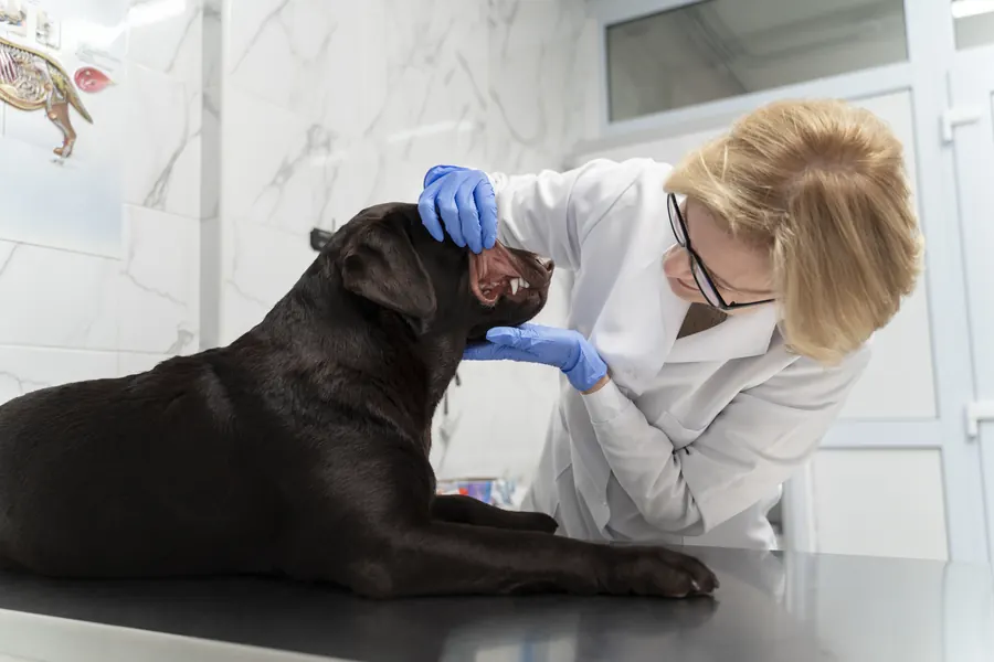
When to see a vet urgently
Have a new lump examined promptly, even if your dog is eating and moving normally.
- Contact a vet promptly if a lesion enlarges, ulcerates or is clearly painful.
- Heavy bleeding requires emergency care.
- If your dog collapses, seek emergency care immediately.
- Difficulty breathing also needs immediate assessment.
Find emergency care through the vet guide.
Overview
Benign and malignant lesions
Skin tumours are among the most common canine tumours. Different tumours can develop from different skin layers and structures. A benign tumour does not spread to other organs. A malignant tumour can invade surrounding tissues.
Of the skin tumours diagnosed at a pathology laboratory in northern Portugal between 2014 and 2020, 62.9% were benign. At the same laboratory, 37.1% were malignant. Mast cell tumours accounted for 22.7% of all these skin tumours. Sebaceous gland tumours accounted for 8.1%.
Benign soft tissue tumours made up 9.7% of skin tumours diagnosed at the same Portuguese laboratory. Of these benign soft tissue tumours, 74.8% were lipomas. Blood vessel tumours include both benign and malignant forms.
Skin tumours also include growths arising from protective surface cells, such as squamous cell carcinoma.
Causes
A lipoma arises from fatty tissue and is often found beneath the skin. A sebaceous tumour develops in a gland whose normal role is to release sebum onto the skin and into the hair follicle. Soft tissue sarcomas include fibrosarcomas arising from connective tissue and peripheral nerve sheath tumours. More rarely, a tumour visible in the skin is a metastasis from elsewhere in the body.
Some cutaneous squamous cell carcinomas are associated with prolonged exposure to ultraviolet sunlight. Radiation more readily damages skin with little pigment and a thin coat. Papillomavirus can cause wart-like tumours, but similar lesions also develop without a virus. Genetics may influence the development of some skin tumours.
Hair follicle cells can give rise to a benign keratoacanthoma, for example. Sebaceous gland hyperplasia can resemble a tumour even though it is an enlargement of the gland. A mast cell tumour arises from mast cells involved in immune defence.
Risk factors
The likelihood of a skin tumour increases with age. However, histiocytomas are common in young dogs under 3.5 years old. Benign sebaceous adenomas, by contrast, are often found in elderly dogs. Lipomas are usually diagnosed in ageing dogs, particularly overweight females.
Breed effects depend on the tumour: mast cell tumours, for example, are more common in Boxers. A sun-damage-associated tumour can develop on pale, sparsely haired abdominal skin. Age or breed cannot establish whether an individual dog's lump is benign or malignant; the tumour needs investigation.
Hyperplasia and tumours of the hepatoid glands around the anus occur around three times as often in uncastrated males as in females. Solitary wart-like skin lesions are more common in elderly dogs. Multiple papillomas on the skin or mucous membranes are typically seen in younger animals.
Typical symptoms
The first finding is often a lump that can be felt or rises above the skin, even if the dog seems unchanged. A skin tumour's surface may be smooth, hairless, crusted or uneven and wart-like.
- A single new nodule may appear on the skin.
- A lesion may feel like a movable lump beneath the skin.
- The surface may ulcerate, remain open or bleed.
- Sometimes the most obvious change is skin discolouration or a hairless patch.
- A lesion may itch, particularly if irritated.
- A toe lesion may cause lameness or loss of a claw.
A histiocytoma is often a pink, hairless lump around 1–2 cm wide. A sebaceous adenoma can develop a crusted surface. A dark skin lesion may be a benign pigment-cell tumour, called a melanocytoma.
A soft red or blackish-red lump may be a haemangioma arising from blood vessels. In the Portuguese laboratory's skin tumour cases, 14.6% had lesions at multiple sites. A hindlimb was recorded as the single location in 12.1% of cases.
Cutaneous lymphoma can appear as flat patches or thickened areas as well as raised lumps. Early lesions may resemble allergic skin disease, for example. In some dogs, lymphoma lesions mainly affect the pads or mucous membranes.
Complications and consequences
A benign lipoma growing in an awkward location can impede movement. A lump on the eyelid margin can irritate the eye's surface if it touches it. A sebaceous adenoma's crusted surface can become infected. A large tumour around the anus can press on it and make defaecation difficult.
A soft tissue sarcoma of the skin or subcutaneous tissue can invade surrounding tissues. The estimated metastatic rate for grade one and two soft tissue sarcomas is around 15%. For grade three tumours, it is around 50%. Metastases from some skin tumours may be found in local lymph nodes or lungs.
A tumour may extend deeper than the visible lump suggests. A cutaneous haemangioma can ulcerate despite being benign. Malignant blood vessel tumours can cause death of neighbouring tissue.
Soft tissue sarcoma metastases may become apparent after a delay: median time to clinically detectable spread is around one year.
Diagnosis at the vet
Examining and sampling the lump
The vet establishes where the lesion is and whether it moves or is attached to deeper tissues. Appearance alone cannot distinguish benign from malignant lesions. A fine-needle sample provides individual cells for microscopic examination. It can often identify a lipoma, cyst or mast cell tumour, for example.
If fine-needle sampling is insufficient for diagnosis, a biopsy can be taken. In a tissue sample, the pathologist sees individual cells and how the tumour is arranged relative to surrounding tissue. Histology can often determine tumour grade. Visible cell divisions can also be counted microscopically in the tissue.
If inflammation is suspected beneath a patch, a skin surface sample may reveal bacteria or yeast. Fine-needle sampling is usually a minor procedure, often requiring neither sedation nor general anaesthesia. A sample from a fatty lump may contain few cells, making interpretation harder.
A biopsy can also be taken from part of a tumour if removing the whole lesion at once is inappropriate. Identifying some tumours requires special tissue stains.
Assessing local growth and metastases
Staging establishes where tumour tissue can be detected. Nearby lymph nodes may be examined according to tumour type. Node size does not reliably indicate whether tumour cells are present. CT or MRI can help plan surgery for a large soft tissue sarcoma or one attached to deeper tissues.
Three-view chest X-rays may be taken when staging a soft tissue sarcoma. Imaging is chosen according to whether information about spread would affect treatment or prognosis. Blood and urine tests help assess general health.
Treatment
Monitoring or removal
The vet prescribes treatment and any medication. Treatment depends on tumour type, size and location, and the dog's general condition. A confirmed benign lesion without symptoms may be monitored if it does not ulcerate or interfere with normal function. Most confirmed canine histiocytomas disappear spontaneously within two to three months. A lipoma impeding movement can be removed despite being benign.
Removing a malignant tumour generally includes some healthy surrounding tissue. The extent of surgery depends partly on how much healthy tissue around it can be removed. The removed lesion is examined microscopically to confirm tumour type. Surgical margins are checked for tumour cells reaching the specimen's edge.
Specimen surfaces can be marked with ink so the pathologist can distinguish true surgical margins from surfaces created during sample handling. Sarcoma removal may aim for lateral margins greater than 3 cm if the location permits. In depth, surgery aims to include the fascial plane beneath the tumour. With benign hepatoid gland adenomas, up to 95% of males may have a complete response to castration.
Additional treatment and local recurrence
If tumour cells remain at a soft tissue sarcoma's surgical margin, the vet may consider re-excising the area. If adequate margins cannot be achieved, radiotherapy may help control local growth. Around 20–35% of incompletely removed soft tissue sarcomas recur locally. Local control of incompletely removed sarcomas subsequently treated with radiotherapy has been 75% at one year. If cancer has spread, the vet may consider systemic chemotherapy according to tumour type.
A narrow margin means tumour cells are close to the cut surface but do not reach the edge. Local recurrence risk may be greater if an incompletely removed soft tissue sarcoma is high-grade.
Prevention
Reducing prolonged, intense sun exposure helps prevent UV-associated squamous cell carcinoma. Avoiding direct sun when radiation is strongest can help protect pale, sparsely haired skin. The exact cause of most cutaneous squamous cell carcinomas is unknown.
Post-treatment visits check for new tumours as well as local recurrence. Follow-up timing depends on the dog's condition, diagnosis and time since treatment.
Spread of contagious papillomavirus can be reduced by keeping an affected dog apart from susceptible dogs. The virus can also spread through shared objects.
Important for dog owners
Record the lump's location, approximate size and when you noticed it. Report bleeding, breakdown of the surface or discomfort. Do not squeeze a lump empty: cyst contents, for example, can spread into surrounding skin and cause inflammation. Monitor the surgical site and report a new lump developing in an old scar.
Mention other skin lesions you have noticed, as one dog can have lumps at several sites. Also show the vet a changed claw if your dog is lame or a toe is swollen.
Frequently asked questions
Can a benign lump be left in place?
Yes, if its type has been established and it does not interfere with movement or other normal function. However, a lipoma in an awkward location may need removal. An ulcerated or painful lesion should be reassessed by the vet.
Does a dark lump mean melanoma?
Dark colour does not identify a tumour, because a benign pigment-cell tumour can also have a dark surface. A malignant melanoma can also be pale. A sample is needed to identify the lesion, as appearance cannot provide a definitive diagnosis.
Why is a biopsy sometimes needed after fine-needle sampling?
A fine-needle sample provides information about the cells and often identifies the lump type. A biopsy allows the pathologist to examine tissue structure and tumour invasion into surrounding tissue. If the entire lump is removed, histology can also assess surgical margins.
What does an incomplete surgical margin mean?
It means tumour cells are visible right up to the edge of the removed tissue. Tumour cells may therefore remain in the dog's skin or underlying tissue. For a soft tissue sarcoma, for example, further surgery or radiotherapy may then be considered to control local growth.
Can one dog have different types of lumps?
Yes: many lesions can occur on and beneath the skin, including lipomas, cysts and sebaceous gland tumours. Have a new lump checked even if a previous lesion was benign. Fine-needle sampling can help identify its type.
Sources
- Tumors of the Skin in Dogs, Merck Veterinary Manual, accessed 2026-11-06.
- Koirien ja kissojen ihomuutokset, Nikulan Eläinklinikka, accessed 2026-11-06.
- Iho- ja korvasairaudet, Eläinlääkäriasema Kamu, accessed 2026-11-06.
- Epidermal and Hair Follicle Tumors in Animals, Merck Veterinary Manual, accessed 2026-11-06.
- Koiran ihomuutosten diagnosointi ja hoito, SoVet, accessed 2026-11-06.
- 2026 Oncology Guidelines: Tumor Diagnostics and Staging, AAHA, accessed 2026-11-06.
- 2026 Oncology Guidelines: Surgery, AAHA, accessed 2026-11-06.
- 2026 Oncology Guidelines: Post-Treatment Monitoring, AAHA, accessed 2026-11-06.
- Emergency and Critical Care: emergent conditions, Cornell University College of Veterinary Medicine, accessed 2026-11-06.
- 2026 Oncology Guidelines: Soft Tissue Sarcoma, AAHA, accessed 2026-11-06.
- Retrospective study of canine cutaneous tumors in Northern Portugal (2014–2020), Companion Animal Health and Genetics, accessed 2026-11-06.
- Lymphocytic, Histiocytic, and Related Cutaneous Tumors in Animals, MSD Veterinary Manual, accessed 2026-11-06.
This information does not replace a visit to a vet. In an emergency, contact your nearest emergency vet: find a vet (RCVS).
More on this topic: Skin and coat
- Acne in dogs: chin spots and treatment
- Alopecia X and seasonal flank alopecia in dogs
- Paw pad injuries in dogs: first aid and wound care
- Atopic dermatitis in dogs: itching, tests and long-term care
- Demodectic mange in dogs: Demodex mites and skin disease
- Fibrosarcoma in dogs: symptoms, diagnosis and treatment