
Dog diseases · Reproductive system
Testicular tumours in dogs – symptoms and treatment
Testicular tumours particularly affect older, uncastrated male dogs. Most are benign. However, some release hormones that can damage the bone marrow.
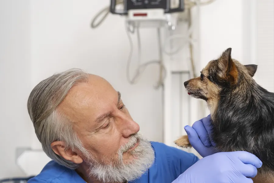
When to see a vet urgently
Hormonal disturbances can cause anaemia or bleeding even when a tumour has not spread.
- Your dog's gums become pale and they are clearly weak.
- Sudden, severe abdominal pain develops; a retained testicle may have twisted.
- Bleeding occurs alongside other signs of illness.
- Urination becomes clearly difficult or no urine is passed at all.
These situations need urgent veterinary assessment. The vet guide can help you find the nearest assistance.
Overview
A testicular tumour is a growth caused by uncontrolled multiplication of cells in a testicle. The most common types are Leydig cell tumours, Sertoli cell tumours and seminomas.
Testicular tumours have been estimated to account for 16–27% of all tumours in male dogs. A proportion of 4–7% has also been reported, so frequency figures differ.
These proportions describe the distribution of tumours, rather than the proportion of all male dogs that develop one. Leydig cell tumours account for at least 50% of testicular tumours in some studied groups of dogs.
Sertoli cell tumours have been reported to account for 7.4–8%. The harm a tumour causes may relate more to hormone secretion than to malignant growth.
Causes
Which cells give rise to the tumour?
A seminoma develops from cells in the sperm-producing cell line. Leydig cells lie between the seminiferous tubules and normally produce male sex hormones.
Sertoli cells support sperm development within the seminiferous tubules. More than one tumour type can occur in the same testicle at once.
A mixed tumour may contain tumour cells from both the sperm-producing cell line and supporting tissue. Other tumour types arising in the testicle are rare.
Growth of a Sertoli cell tumour can compress and destroy surrounding normal testicular tissue.
Why do hormonal signs develop?
More than 50% of Sertoli cell tumours produce oestrogens, the female sex hormones. About 70% of Sertoli cell tumours in the abdomen are hormonally active.
Leydig cell tumours produce hormones that cause symptoms much less often. The precise cause of a tumour often remains unclear, and several genetic and environmental factors may contribute.
Oestrogen from the tumour can suppress normal hormonal control by the brain and pituitary gland.
Risk factors
Testicular tumours are particularly diagnosed in dogs over ten years old. Younger dogs can also be affected, especially if they have a retained testicle.
The tumour risk is particularly important in cryptorchid males over six years old. A retained testicle is one that has remained in the abdomen or groin instead of the scrotum.
The tumour risk in a retained testicle has been estimated at about 13 times that in a normally descended testicle. Cryptorchidism particularly predisposes dogs to Sertoli cell tumours and seminomas, but no equivalent association with Leydig cell tumours has been found.
Breed associations have been described in Boxers, German Shepherds and Afghan Hounds, for example. A tumour can also develop in a testicle that descended completely normally into the scrotum.
Bilateral involvement has been reported in 43% of Leydig cell tumours, 11% of Sertoli cell tumours and 18% of seminomas. Both testicles are therefore examined even if a visible change affects only one side.
Typical symptoms
Many tumours are found by chance without visible signs of illness.
- A new lump or uneven thickening can be felt in a testicle.
- The testicles have become different sizes or the scrotum swells.
- The male dog's nipples or mammary glands enlarge.
- Hair is lost symmetrically from both sides of the body.
- Skin darkens in hairless areas.
- The sheath of the penis hangs down more than usual.
- Other males show more interest in the dog than usual.
- Sexual interest decreases or the dog begins squatting to urinate.
The other testicle may also shrink because of hormonal changes.
Disease that has spread may cause weight loss, poor appetite or vomiting.
Difficulty urinating or passing stools may result from metastases near the urinary tract or prostate.
Hormonal signs from a Sertoli cell tumour may also include milk production from the male's mammary glands.
Hormone-related hairless areas can appear around the genitals, inner thighs, abdomen, chest and shoulder blades.
Complications and consequences
Bone marrow damage and spread
Excess oestrogen can reduce blood cell production in the bone marrow. A fall in red blood cells causes anaemia, which may appear as pallor and tiredness.
A reduced platelet count can cause bleeding. Infection-fighting white blood cells may also decrease.
Hair loss, anaemia or bone marrow damage has been described in up to 15% of dogs with an oestrogen-secreting Sertoli cell tumour. The reported risk of spread for Sertoli cell tumours and seminomas is below 15%.
More specific estimates are 0.6–9.0% for Sertoli cell tumours and below 10% for seminomas. Leydig cell tumours only rarely metastasise.
Metastases may affect abdominal lymph nodes and organs such as the lungs. The outlook is generally good if the tumour is removed and the dog has neither metastases nor bone marrow damage.
Hormonal effects may cause changes in the prostate's lining cells and cystic enlargement. A larger Sertoli cell tumour may have a greater risk of spread.
Diagnosis at the vet
Examination and imaging
The vet palpates both testicles and assesses their size and structure. A testicle missing from the scrotum raises suspicion of cryptorchidism.
Abdominal ultrasound is used to look for a retained testicle or abdominal tumour. Ultrasound can also assess changes to a testicle's internal structure caused by a tumour.
Tests to assess spread may include blood tests, urinalysis and chest X-rays. Rectal palpation adds to the examination of the pelvic area.
CT may be needed to assess abdominal lymph nodes and organs in a cryptorchid dog. An enlarged or abnormal-feeling lymph node may be sampled to investigate metastasis.
Hormones and tissue samples
Investigation of hormonal signs may include measuring testosterone and oestradiol and assessing their ratio. A cell sample from the penile sheath may show the effects of oestrogen on the tissue.
The definitive tumour type is usually confirmed by microscopic examination of the removed testicle, called histopathology. The pathologist also assesses tissue features related to the tumour's behaviour.
In particular circumstances, a fine-needle sample may establish whether a lesion is a tumour or, for example, a cyst. However, a needle sample or biopsy can damage the protective blood–testis barrier and reduce fertility.
Seminoma cells may look malignant under the microscope even when the tumour behaves benignly.
Why is tissue examination needed?
A ten-year review of biopsies in the United States examined 156 testicles from 107 dogs. A tumour was found in 92 testicles, or 59%.
A non-tumour lesion was found in 46 testicles, or 29% of those examined. These other findings included inflammation, circulatory disturbance and testicular shrinkage.
No significant microscopic abnormality was found in 13 testicles, or 8%. More than 15% contained multiple tumours within the same testicle.
These were biopsies submitted for examination, so the proportions do not describe the disease risk for all dogs. When needed, immunohistochemical stains help the pathology laboratory refine tumour classification.
Cancer arising elsewhere in the urinary or reproductive system can also metastasise to the testicle. Extensive connective tissue in a Sertoli cell tumour may make it particularly firm.
A Leydig cell tumour may appear on tissue examination as a soft, yellow nodule containing haemorrhage. A seminoma can become extensive enough to enlarge the whole testicle or markedly distort its shape.
Treatment
Surgery and choosing further treatment
The most common treatment is surgical removal of the testicles, or castration. Removing a retained testicle and a hormone-secreting tumour is particularly important to prevent complications.
A retained testicle in the groin can usually be removed through a skin incision over it. An abdominal testicle is removed through an operation accessing the abdomen.
Removal of only the affected testicle may be considered if the other appears normal on examination and ultrasound, and fertility is to be preserved. Blood results and hormone function need monitoring after a hormonally active tumour.
Chemotherapy or radiotherapy may be considered for disease that has spread. The vet prescribes postoperative pain relief and reviews the need for it during recovery.
Recovery at home
Prevent licking of the surgical site with the collar or other suitable protection recommended by the vet. Keep the wound dry and do not allow swimming until it has healed.
Restrict running, jumping, rough play and stair use according to the discharge instructions. Initially, outings are on a lead for toilet breaks.
Appetite may be reduced during the first 24 hours after anaesthesia. Mild bruising or redness can be part of normal wound healing during the first few days.
Persistent discharge, pus or a bad smell needs reassessment.
Do not put creams, sprays or other topical products on the wound without the treating vet's explicit instructions. Quiet activities, such as food puzzles, can help during recovery while exercise is restricted.
Do not change the diet abruptly during recovery without specific veterinary advice. Passing stools may be delayed for 1–2 days after surgery, partly because of reduced food intake.
Increase activity gradually according to wound healing and follow-up findings before returning to normal walks.
Keep fresh water available unless the vet has given specific instructions for the situation.
Prevention
Removing the testicles prevents tumours from developing in them later. When assessing the overall benefits of castration, consider that many factors besides sex hormones affect life expectancy.
Removal of both testicles is recommended for a cryptorchid dog because of tumour and torsion risks. A cryptorchid dog should not be bred from because failure of descent is inherited.
If the other testicle is preserved during tumour removal, a similar tumour may develop in it later. A health examination of an uncastrated male can identify a change before obvious symptoms develop.
Important for dog owners
Note when you first noticed a lump or size difference and any changes in the coat, nipples and behaviour. Tell the practice whether both testicles were previously in the scrotum. Bring records of earlier examinations and operations.
An enlarged testicle may also result from inflammation or another non-tumour change.
After surgery, ask for the histopathology result and a written follow-up plan. Establish whether both testicles were removed, whether blood tests are needed and which findings require another visit. Take photographs during healing if wound changes are difficult to describe.
Visible external skin stitches or staples are removed by a veterinary professional at the practice.
Dissolving stitches beneath the skin do not usually need removal.
Contact the practice before the scheduled check if pain or weakness worsens.
Frequently asked questions
Are testicular tumours in dogs always cancerous?
No. Most testicular tumours are benign. However, even a benign tumour can release hormones and disrupt blood cell production. The type is usually confirmed by tissue examination after surgical removal. Leydig cell tumours only rarely spread to lymph nodes or other organs.
Can a dog develop a tumour if both testicles have descended?
Yes. Sertoli cell tumours have also been found in testicles within the scrotum. Cryptorchidism increases the risk, but a descended testicle is not protected from a tumour. A new difference in testicle size or a lump is therefore a reason for examination. Many dogs show no obvious signs of illness at all.
Why are my male dog's nipples enlarging and his hair falling out?
An oestrogen-secreting Sertoli cell tumour can cause mammary gland enlargement and hair loss. Other males may also become interested in the dog. Excess oestrogen can affect the bone marrow too, so these signs need investigation. Anaemia may cause pale gums and weakness.
Must both testicles be removed?
Removal of both testicles is the usual treatment. In selected cases, the other testicle may be preserved if it appears normal on examination and ultrasound and fertility is to be retained. However, a tumour may develop in it later. Blood results are also monitored after a hormonally active tumour.
Can a testicular tumour spread elsewhere in the body?
Yes, but most have a low risk of spread. The reported risk for Sertoli cell tumours and seminomas is below 15%. A suspicious lymph node may be sampled so that further treatment can be planned according to the findings. Spread of Leydig cell tumours is particularly rare.
Sources
- WSAVA guidelines for the control of reproduction in dogs and cats (2024), World Small Animal Veterinary Association, accessed 2026-11-03.
- Testicular Tumors, VCA Animal Hospitals, accessed 2026-11-03.
- Testicular Tumors, Veterinary Society of Surgical Oncology, accessed 2026-11-03.
- Koirani toinen kives ei ole laskeutunut. Onko sillä kohonnut syöpäriski?, FirstVet, accessed 2026-11-03.
- Canine Testicular Neoplasia, MSPCA-Angell, accessed 2026-11-03.
- Clinical Management of Testicular Tumors in Dogs (2026): abstract and bibliographic record, Animals / PubMed, accessed 2026-11-03.
- Cryptorchidism in dogs (retained testicle), Cornell University College of Veterinary Medicine, accessed 2026-11-03.
- Postoperative Care After Surgery: What Animal Owners Should Expect, American College of Veterinary Surgeons, accessed 2026-11-03.
This information does not replace a visit to a vet. In an emergency, contact your nearest emergency vet: find a vet (RCVS).
More on this topic: Reproductive system
- Vaginitis in dogs: discharge, symptoms and treatment
- Vaginal hyperplasia in dogs: swelling and treatment
- Balanoposthitis in dogs – discharge and inflammation
- Enlarged prostate in dogs – symptoms and treatment
- Prostatitis in dogs: prostate infection and treatment
- Infertility in dogs – causes, investigations and treatment